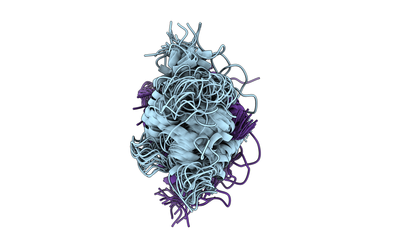
1UWO image
Deposition Date 1997-12-05
Release Date 1998-06-10
Last Version Date 2024-05-22
Entry Detail
PDB ID:
1UWO
Title:
CALCIUM FORM OF HUMAN S100B, NMR, 20 STRUCTURES
Biological Source:
Source Organism:
Homo sapiens (Taxon ID: 9606)
Host Organism:
Method Details:
Experimental Method:
Conformers Calculated:
30
Conformers Submitted:
20
Selection Criteria:
LEAST RESTRAINT VIOLATIONS
Macromolecular Entities
Structures with similar UniProt ID
Protein Blast
Polymer Type:polypeptide(L)
Molecule:S100B
Gene (Uniprot):S100B
Chain IDs:A, B
Chain Length:91
Number of Molecules:2
Biological Source:Homo sapiens
Ligand Molecules
Primary Citation
A novel calcium-sensitive switch revealed by the structure of human S100B in the calcium-bound form.
Structure 6 211 222 (1998)
PMID: 9519411 DOI: 10.1016/S0969-2126(98)00022-7

Abstact

BACKGROUND S100B is a homodimeric member of the EF-hand calcium-binding protein superfamily. The protein has been implicated in cellular processes such as cell differentiation and growth, plays a role in cytoskeletal structure and function, and may have a role in neuropathological diseases, such as Alzheimers. The effects of S100B are mediated via its interaction with target proteins. While several studies have suggested that this interaction is propagated through a calcium-induced conformational change, leading to the exposure of a hydrophobic region of S100B, the molecular details behind this structural alteration remain unclear. RESULTS The solution structure of calcium-saturated human S100B (Ca(2+)-S100B) has been determined by heteronuclear NMR spectroscopy. Ca(2+)-S100B forms a well defined globular structure comprising four EF-hand calcium-binding sites and an extensive hydrophobic dimer interface. A comparison of Ca(2+)-S100B with apo S100B and Ca(2+)-calbindin D9k indicates that while calcium-binding to S100B results in little change in the site I EF-hand, it induces a backbone reorientation of the N terminus of the site II EF-hand. This reorientation leads to a dramatic change in the position of helix III relative to the other helices. CONCLUSIONS The calcium-induced reorientation of calcium-binding site II results in the increased exposure of several hydrophobic residues in helix IV and the linker region. While following the general mechanism of calcium modulatory proteins, whereby a hydrophobic target site is exposed, the 'calcium switch' observed in S100B appears to be unique from that of other EF-hand proteins and may provide insights into target specificity among calcium modulatory proteins.

Legend

Protein

Chemical

Disease

Primary Citation of related structures