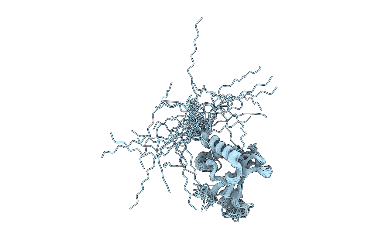
1UW0 image
Entry Detail
PDB ID:
1UW0
Keywords:
Title:
Solution structure of the zinc-finger domain from DNA ligase IIIa
Biological Source:
Source Organism:
Host Organism:
PDB Version:
Deposition Date:
2004-01-27
Release Date:
2004-08-05
Method Details:
Experimental Method:
Conformers Calculated:
50
Conformers Submitted:
28
Selection Criteria:
LOW RESTRAINT VIOLATIONS
Macromolecular Entities
Polymer Type:polypeptide(L)
Description:DNA LIGASE III
Chain IDs:A
Chain Length:117
Number of Molecules:1
Biological Source:HOMO SAPIENS
Ligand Molecules
Primary Citation
Solution Structure and DNA Binding of the Zinc-Finger Domain from DNA Ligase Iiialpha
J.Mol.Biol. 341 723 ? (2004)
PMID: 15288782 DOI: 10.1016/J.JMB.2004.06.035

Abstact

DNA ligase IIIalpha carries out the final ligation step in the base excision repair (BER) and single strand break repair (SSBR) mechanisms of DNA repair. The enzyme recognises single-strand nicks and other damage features in double-stranded DNA, both through the catalytic domain and an N-terminal domain containing a single zinc finger. The latter is homologous to other zinc fingers that recognise damaged DNA, two in the N terminus of poly(adenosine-ribose)polymerase and three in the N terminus of the Arabidopsis thaliana nick-sensing DNA 3'-phosphoesterase. Here, we present the solution structure of the zinc-finger domain of human DNA ligase IIIalpha, the first structure of a finger from this group. It is related to that of the erythroid transcription factor GATA-1, but has an additional N-terminal beta-strand and C-terminal alpha-helix. Chemical shift mapping using a DNA ligand containing a single-stranded break showed that the DNA-binding surface of the DNA-ligase IIIalpha zinc finger is substantially different from that of GATA-1, consistent with the fact that the two proteins recognise very different features in the DNA. Likely implications for DNA binding are discussed.

Legend

Protein

Chemical

Disease

Primary Citation of related structures