1UV7 image
Entry Detail
PDB ID:
1UV7
Keywords:
Title:
periplasmic domain of EpsM from Vibrio cholerae
Biological Source:
Source Organism:
Host Organism:
PDB Version:
Deposition Date:
2004-01-15
Release Date:
2004-04-23
Method Details:
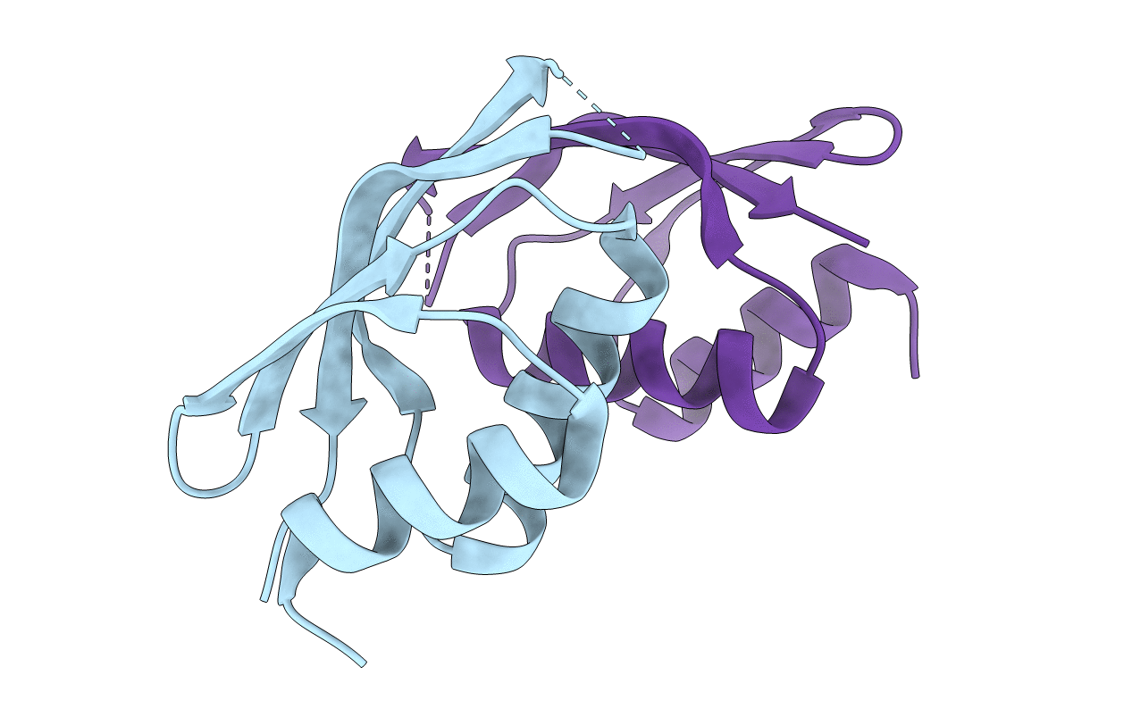
Experimental Method:
Resolution:
1.70 Å
R-Value Free:
0.24
R-Value Work:
0.19
Space Group:
P 32 2 1
Macromolecular Entities
Polymer Type:polypeptide(L)
Description:GENERAL SECRETION PATHWAY PROTEIN M
Chain IDs:A, B
Chain Length:110
Number of Molecules:2
Biological Source:VIBRIO CHOLERAE
Modified Residue
Compound ID Chain ID Parent Comp ID Details 2D Image
MSE A MET SELENOMETHIONINE
Primary Citation
The Crystal Structure of the Periplasmic Domain of the Type II Secretion System Protein Epsm from Vibrio Cholerae: The Simplest Version of the Ferredoxin Fold
J.Mol.Biol. 338 585 ? (2004)
PMID: 15081815 DOI: 10.1016/J.JMB.2004.01.064

Abstact

The terminal branch of the general secretion pathway (Gsp or type II secretion system) is used by several pathogenic bacteria for the secretion of their virulence factors across the outer membrane. In these secretion systems, a complex of 12-15 Gsp proteins spans from the pore in the outer membrane via several associated signal or energy-transducing proteins in the inner membrane to a regulating ATPase in the cytosol. The human pathogen Vibrio cholerae uses such a system, called the Eps system, for the export of the cholera toxin and other virulence factors from its periplasm into the lumen of the gastrointestinal tract of the host. Here, we report the atomic structure of the periplasmic domain of the EpsM protein from V.cholerae, which is a part of the interface between the regulating part and the rest of the Eps system. The crystal structure was determined by Se-Met MAD phasing and the model was refined to 1.7A resolution. The monomer consists of two alphabetabeta-subdomains forming a sandwich of two alpha-helices and a four-stranded antiparallel beta-sheet. In the dimer, a deep cleft with a polar rim and a hydrophobic bottom made by conserved residues is located between the monomers. This cleft contains an extra electron density suggesting that this region might serve as a binding site of an unknown ligand or part of a protein partner. Unexpectedly, the fold of the periplasmic domain of EpsM is an undescribed circular permutation of the ferredoxin fold.

Legend

Protein

Chemical

Disease

Primary Citation of related structures