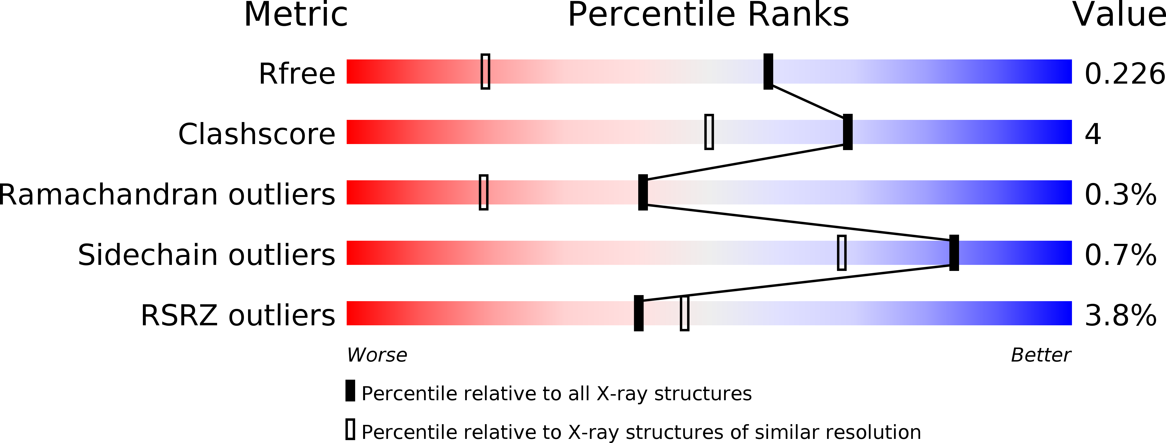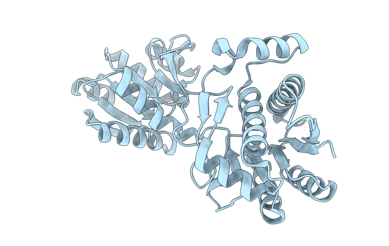
1USG image
Deposition Date 2003-11-24
Release Date 2003-12-18
Last Version Date 2024-10-16
Entry Detail
PDB ID:
1USG
Title:
L-leucine-binding protein, apo form
Biological Source:
Source Organism:
ESCHERICHIA COLI (Taxon ID: 562)
Host Organism:
Method Details:
Experimental Method:
Resolution:
1.53 Å
R-Value Free:
0.21
R-Value Work:
0.18
R-Value Observed:
0.19
Space Group:
C 1 2 1
Macromolecular Entities
Polymer Type:polypeptide(L)
Molecule:LEUCINE-SPECIFIC BINDING PROTEIN
Gene (Uniprot):livK
Chain IDs:A
Chain Length:346
Number of Molecules:1
Biological Source:ESCHERICHIA COLI
Primary Citation
X-Ray Structures of the Leucine-Binding Protein Illustrate Conformational Changes and the Basis of Ligand Specificity
J.Biol.Chem. 279 8747 ? (2004)
PMID: 14672931 DOI: 10.1074/JBC.M311890200

Abstact

The periplasmic leucine-binding protein is the primary receptor for the leucine transport system in Escherichia coli. We report here the structure of an open ligand-free form solved by molecular replacement and refined at 1.5-A resolution. In addition, two closed ligand-bound structures of the same protein are presented, a phenylalanine-bound form at 1.8 A and a leucine-bound structure at a nominal resolution of 2.4 A. These structures show the basis of this protein's ligand specificity, as well as illustrating the conformational changes that are associated with ligand binding. Comparison with earlier structures provides further information about solution conformations, as well as the different specificity of the closely related leucine/isoleucine/valine-binding protein.

Legend

Protein

Chemical

Disease

Primary Citation of related structures