1URX image
Deposition Date 2003-11-12
Release Date 2004-03-04
Last Version Date 2023-12-13
Entry Detail
PDB ID:
1URX
Keywords:
Title:
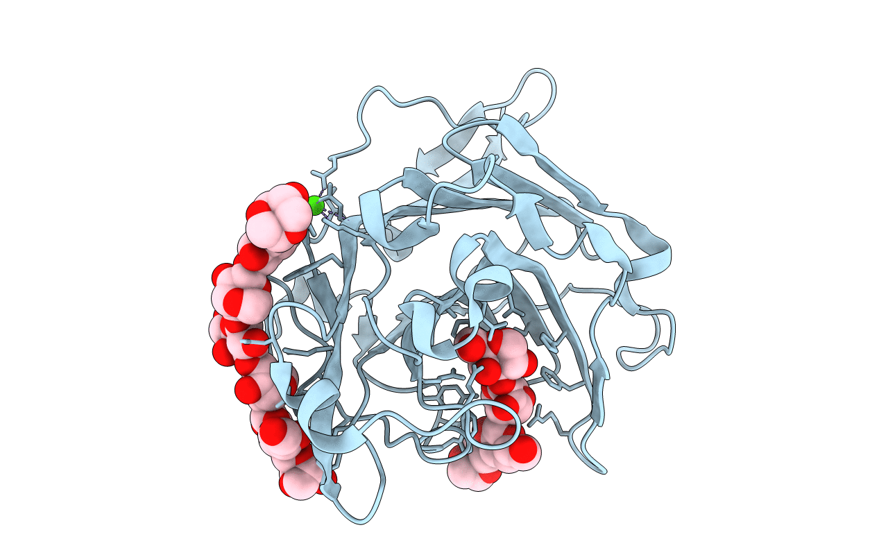
Crystallographic structure of beta-agarase A in complex with oligoagarose
Biological Source:
Source Organism:
Host Organism:
Method Details:
Experimental Method:
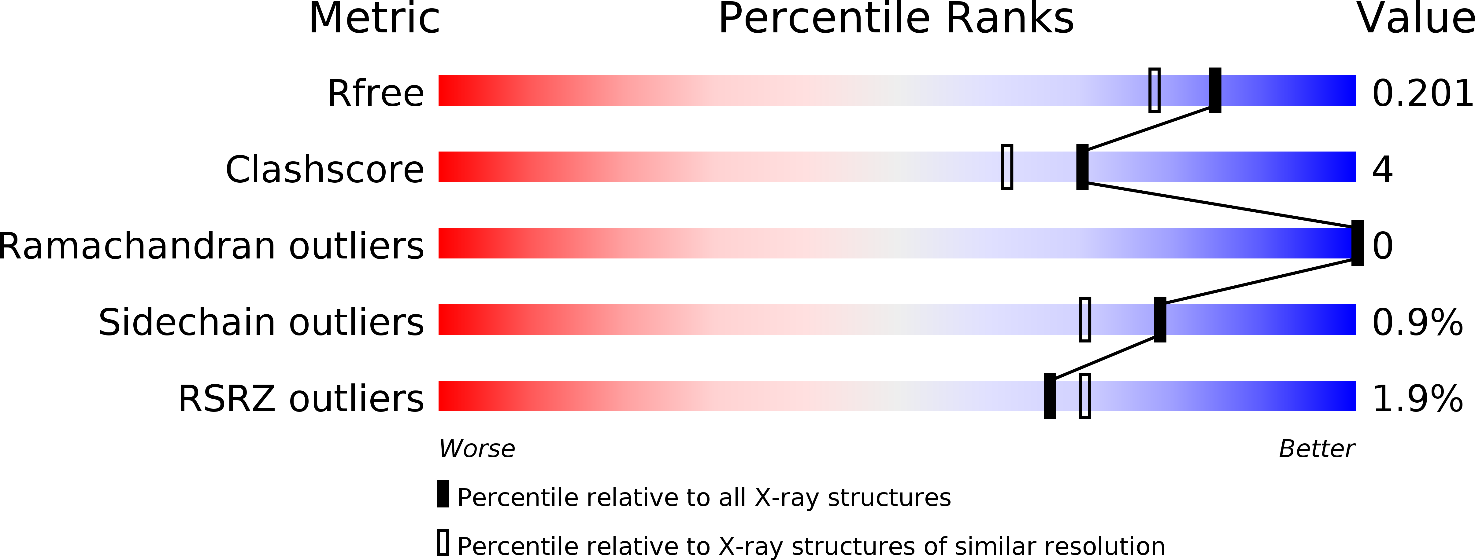
Resolution:
1.70 Å
R-Value Free:
0.18
R-Value Work:
0.15
R-Value Observed:
0.15
Space Group:
P 32 2 1
Macromolecular Entities
Structures with similar UniProt ID
Protein Blast
Polymer Type:polypeptide(L)
Molecule:BETA-AGARASE A
Mutagens:YES
Chain IDs:A
Chain Length:282
Number of Molecules:1
Biological Source:ZOBELLIA GALACTANIVORANS
Ligand Molecules
Primary Citation
Parallel Substrate Binding Sites in a Beta-Agarase Suggest a Novel Mode of Action on Double-Helical Agarose
Structure 12 623 ? (2004)
PMID: 15062085 DOI: 10.1016/J.STR.2004.02.020

Abstact

Agarose is a gel-forming polysaccharide with an alpha-L(1,4)-3,6-anhydro-galactose, beta-D(1,3)-galactose repeat unit, from the cell walls of marine red algae. beta-agarase A, from the Gram-negative bacterium Zobellia galactanivorans, is secreted to the external medium and degrades agarose with an endo-mechanism. The structure of the inactive mutant beta-agarase A-E147S in complex with agaro-octaose has been solved at 1.7 A resolution. Two oligosaccharide chains are bound to the protein. The first one resides in the active site channel, spanning subsites -4 to -1. A second oligosaccharide binding site, on the opposite side of the protein, was filled with eight sugar units, parallel to the active site. The crystal structure of the beta-agarase A with agaro-octaose provides detailed information on agarose recognition in the catalytic site. The presence of the second, parallel, binding site suggests that the enzyme might be able to unwind the double-helical structure of agarose prior to the catalytic cleavage.

Legend

Protein

Chemical

Disease

Primary Citation of related structures
Feedback Form
Name
Email
Institute
Feedback