1UPN image
Deposition Date 2003-10-08
Release Date 2004-01-07
Last Version Date 2024-10-09
Entry Detail
PDB ID:
1UPN
Keywords:
Title:
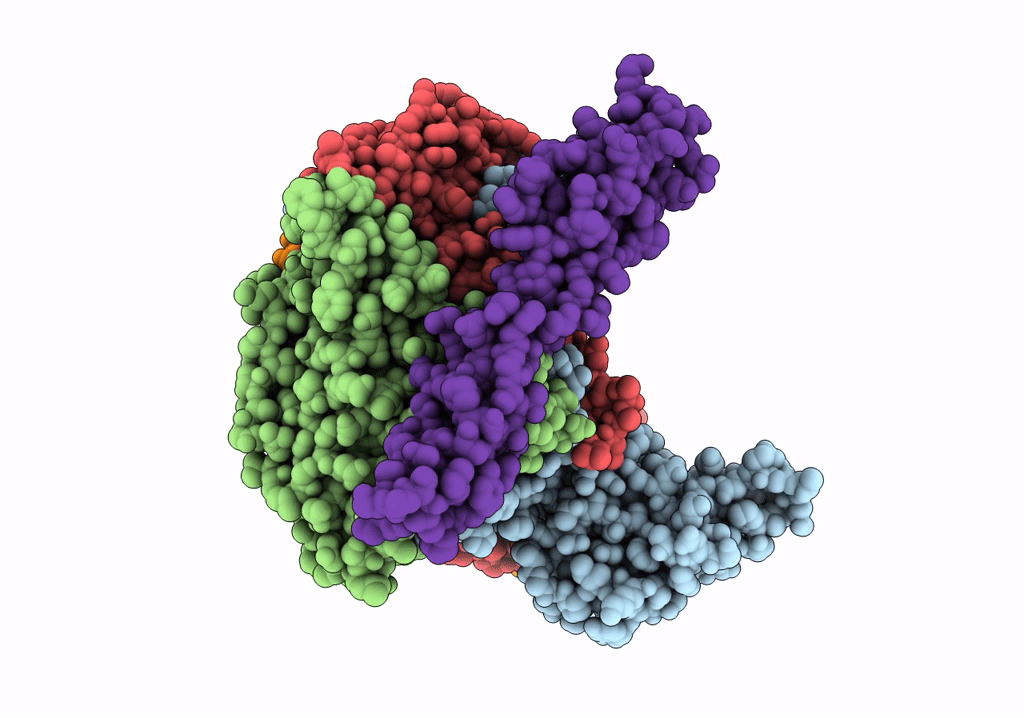
COMPLEX OF ECHOVIRUS TYPE 12 WITH DOMAINS 3 AND 4 OF ITS RECEPTOR DECAY ACCELERATING FACTOR (CD55) BY CRYO ELECTRON MICROSCOPY AT 16 A
Biological Source:
Source Organism:
HOMO SAPIENS (Taxon ID: 9606)
HUMAN ECHOVIRUS 11 (Taxon ID: 12078)
Host Organism:
Method Details:
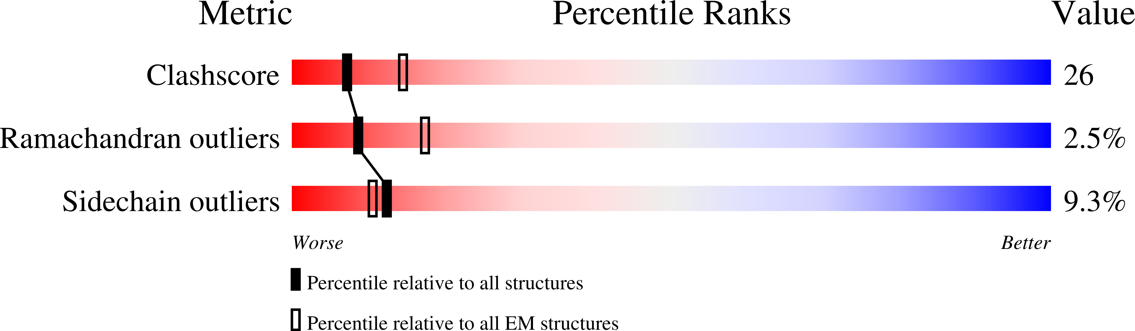
Experimental Method:
Resolution:
16.00 Å
Aggregation State:
PARTICLE
Reconstruction Method:
SINGLE PARTICLE
Macromolecular Entities
Polymer Type:polypeptide(L)
Molecule:ECHOVIRUS 11 COAT PROTEIN VP1
Chain IDs:A
Chain Length:292
Number of Molecules:1
Biological Source:HUMAN ECHOVIRUS 11
Polymer Type:polypeptide(L)
Molecule:ECHOVIRUS 11 COAT PROTEIN VP2
Chain IDs:B
Chain Length:262
Number of Molecules:1
Biological Source:HUMAN ECHOVIRUS 11
Polymer Type:polypeptide(L)
Molecule:ECHOVIRUS 11 COAT PROTEIN VP3
Chain IDs:C
Chain Length:238
Number of Molecules:1
Biological Source:HUMAN ECHOVIRUS 11
Polymer Type:polypeptide(L)
Molecule:ECHOVIRUS 11 COAT PROTEIN VP4
Chain IDs:D
Chain Length:69
Number of Molecules:1
Biological Source:HUMAN ECHOVIRUS 11
Polymer Type:polypeptide(L)
Molecule:COMPLEMENT DECAY-ACCELERATING FACTOR
Gene (Uniprot):CD55
Chain IDs:E
Chain Length:129
Number of Molecules:1
Biological Source:HOMO SAPIENS
Ligand Molecules
Primary Citation
The Structure of Echovirus Type 12 Bound to a Two-Domain Fragment of its Cellular Attachment Protein Decay-Accelerating Factor (Cd 55)
J.Biol.Chem. 279 8325 ? (2004)
PMID: 14634014 DOI: 10.1074/JBC.M311334200

Abstact

Echovirus type 12 (EV12), an Enterovirus of the Picornaviridae family, uses the complement regulator decay-accelerating factor (DAF, CD55) as a cellular receptor. We have calculated a three-dimensional reconstruction of EV12 bound to a fragment of DAF consisting of short consensus repeat domains 3 and 4 from cryo-negative stain electron microscopy data (EMD code 1057). This shows that, as for an earlier reconstruction of the related echovirus type 7 bound to DAF, attachment is not within the viral canyon but occurs close to the 2-fold symmetry axes. Despite this general similarity our reconstruction reveals a receptor interaction that is quite different from that observed for EV7. Fitting of the crystallographic co-ordinates for DAF(34) and EV11 into the reconstruction shows a close agreement between the crystal structure of the receptor fragment and the density for the virus-bound receptor, allowing unambiguous positioning of the receptor with respect to the virion (PDB code 1UPN). Our finding that the mode of virus-receptor interaction in EV12 is distinct from that seen for EV7 raises interesting questions regarding the evolution and biological significance of the DAF binding phenotype in these viruses.

Legend

Protein

Chemical

Disease

Primary Citation of related structures