Deposition Date
2003-09-26
Release Date
2004-11-18
Last Version Date
2024-10-23
Entry Detail
PDB ID:
1UP2
Keywords:
Title:
Structure of the endoglucanase Cel6 from Mycobacterium tuberculosis in complex with glucose-isofagomine at 1.9 angstrom
Biological Source:
Source Organism(s):
MYCOBACTERIUM TUBERCULOSIS (Taxon ID: 83332)
Expression System(s):
Method Details:
Experimental Method:
Resolution:
1.90 Å
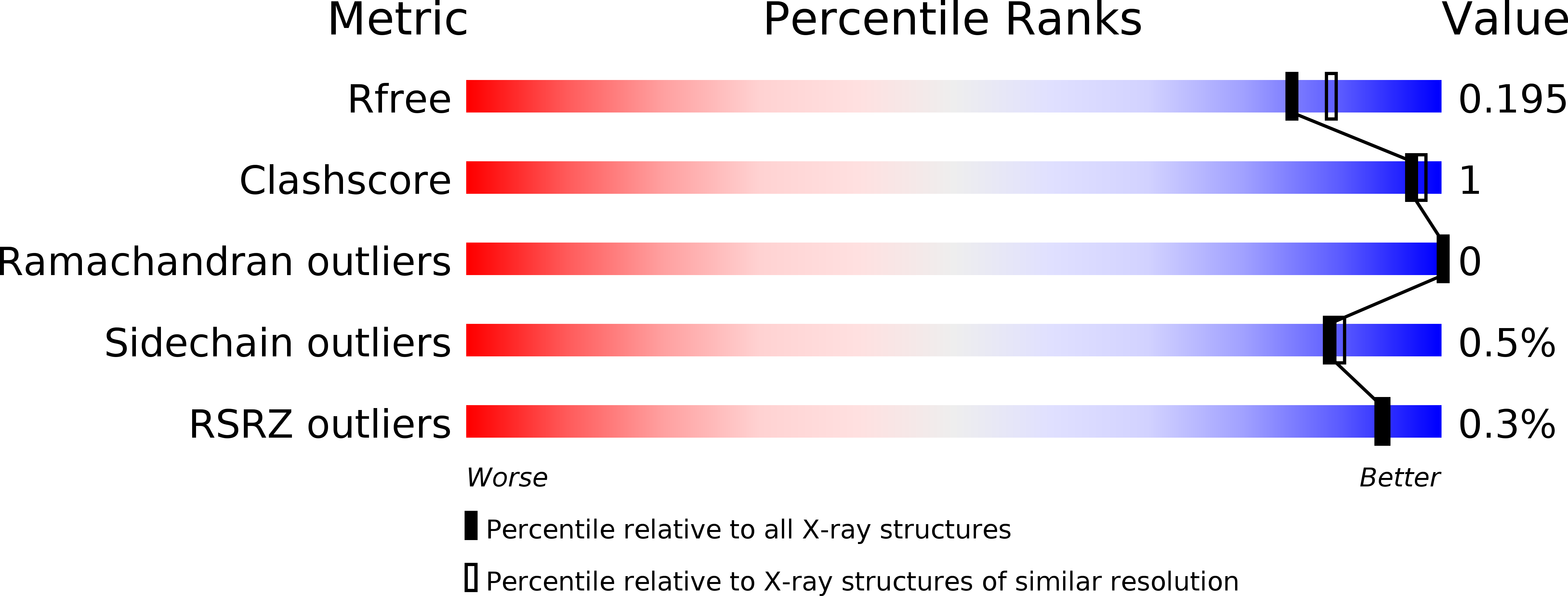
R-Value Free:
0.17
R-Value Work:
0.13
R-Value Observed:
0.13
Space Group:
P 21 21 2