Deposition Date
2003-09-22
Release Date
2003-10-14
Last Version Date
2024-11-20
Entry Detail
PDB ID:
1UOS
Keywords:
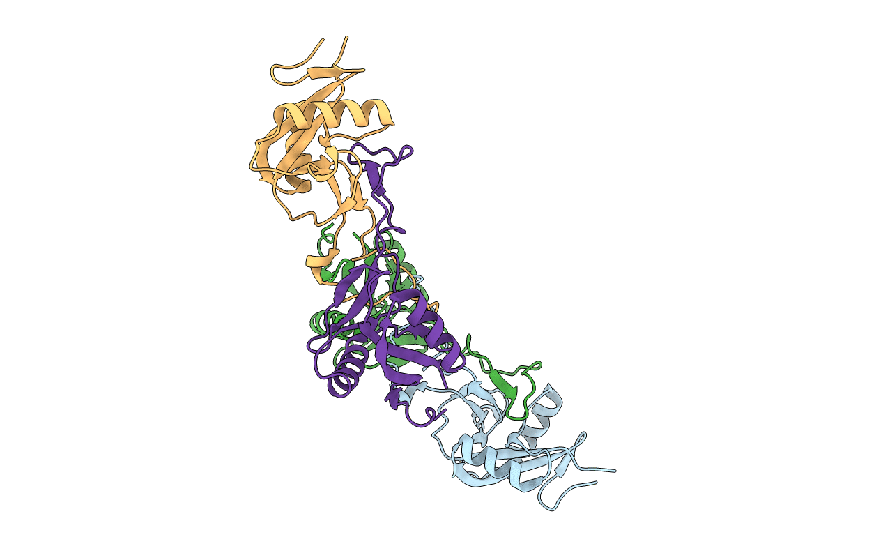
Title:
The Crystal Structure of the Snake Venom Toxin Convulxin
Biological Source:
Source Organism(s):
CROTALUS DURISSUS TERRIFICUS (Taxon ID: 8732)
Method Details:
Experimental Method:
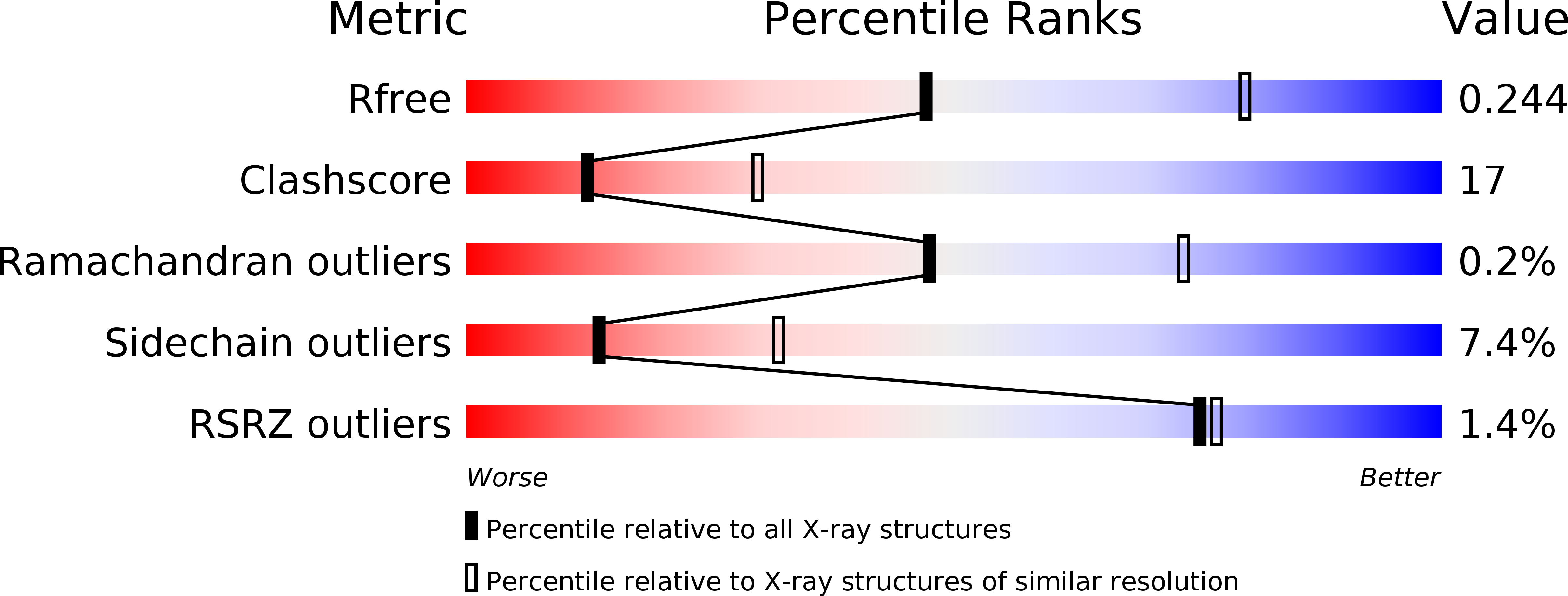
Resolution:
2.70 Å
R-Value Free:
0.26
R-Value Work:
0.23
R-Value Observed:
0.23
Space Group:
I 4