Deposition Date
2003-09-22
Release Date
2003-10-02
Last Version Date
2023-12-13
Entry Detail
PDB ID:
1UOP
Keywords:
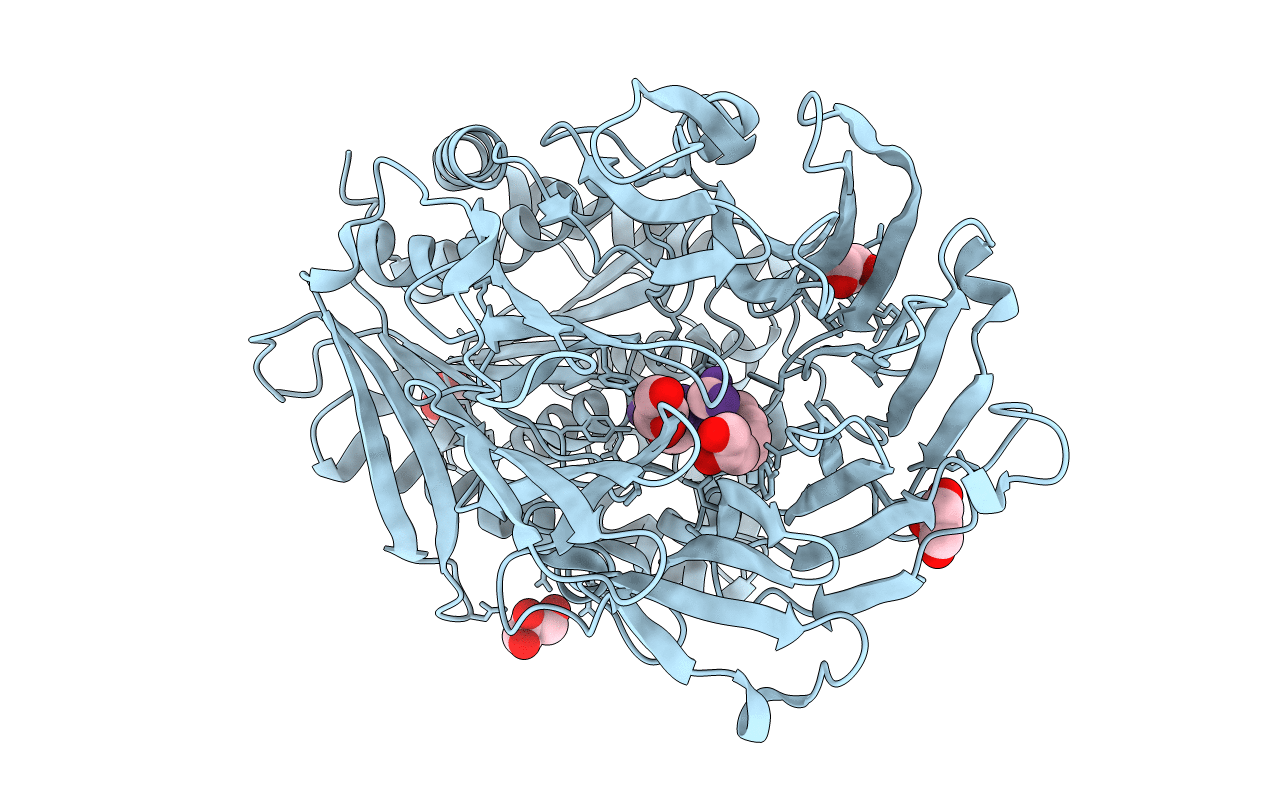
Title:
PROLYL OLIGOPEPTIDASE FROM PORCINE BRAIN, S554A MUTANT WITH BOUND PEPTIDE LIGAND GLY-PHE-GLU-PRO
Biological Source:
Source Organism(s):
SUS SCROFA (Taxon ID: 9823)
synthetic construct (Taxon ID: 32630)
synthetic construct (Taxon ID: 32630)
Expression System(s):
Method Details:
Experimental Method:
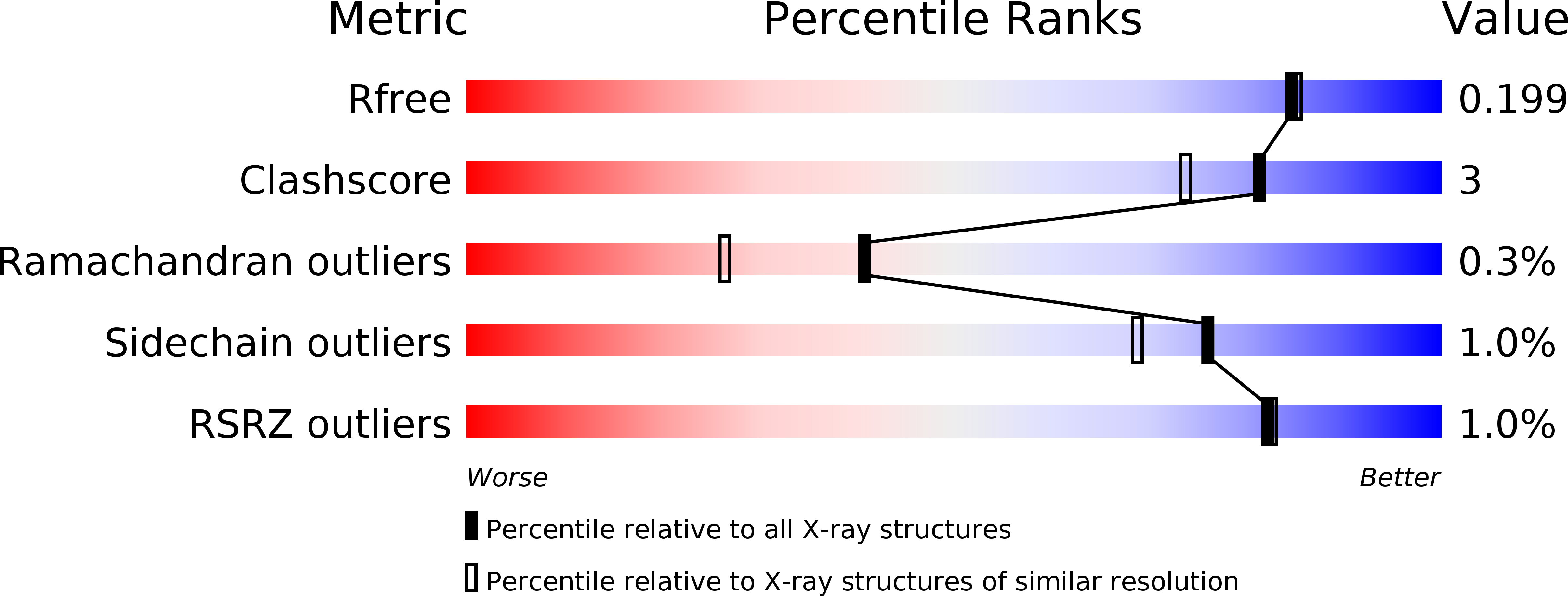
Resolution:
1.85 Å
R-Value Free:
0.19
R-Value Work:
0.15
R-Value Observed:
0.15
Space Group:
P 21 21 21