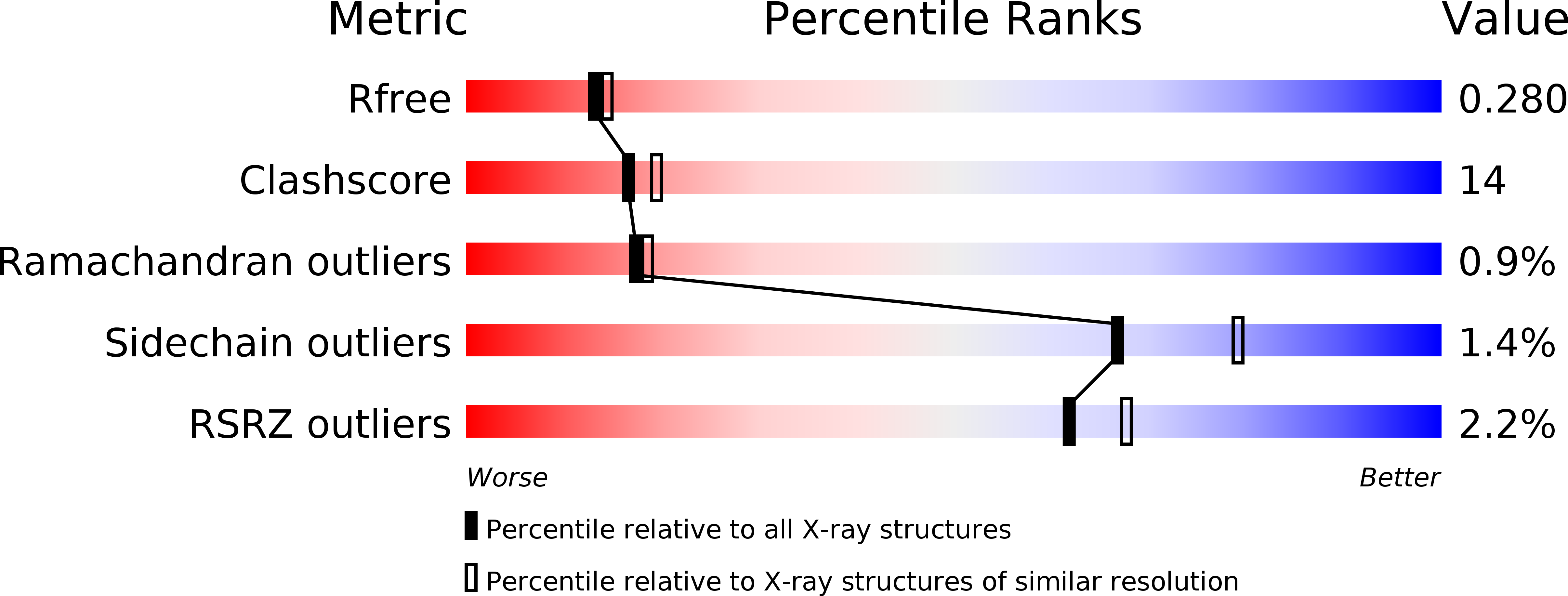
1UOM image
Deposition Date 2003-04-11
Release Date 2003-07-03
Last Version Date 2023-12-13
Entry Detail
PDB ID:
1UOM
Keywords:
Title:
The Structure of Estrogen Receptor in Complex with a Selective and Potent Tetrahydroisochiolin Ligand.
Biological Source:
Source Organism:
HOMO SAPIENS (Taxon ID: 9606)
Host Organism:
Method Details:
Experimental Method:
Resolution:
2.28 Å
R-Value Free:
0.28
R-Value Work:
0.23
Space Group:
P 65 2 2
Macromolecular Entities
Polymer Type:polypeptide(L)
Molecule:ESTROGEN RECEPTOR
Gene (Uniprot):ESR1
Mutations:YES
Chain IDs:A
Chain Length:254
Number of Molecules:1
Biological Source:HOMO SAPIENS
Ligand Molecules
Primary Citation
Estrogen Receptor Modulators: Identification and Structure-Activity Relationships of Potent Eralpha-Selective Tetrahydroisoquinoline Ligands
J.Med.Chem. 46 2945 ? (2003)
PMID: 12825935 DOI: 10.1021/JM030086H

Abstact

As part of a program aimed at the development of selective estrogen receptor modulators (SERMs), tetrahydroisoquinoline derivative 27 was discovered by high throughput screening. Successive replacements of the p-F substituent of 27 by an aminoethoxy side chain and of the 1-H of the tetrahydroisoquinoline core by a 1-Me group provided analogues 19 and 20. These compounds showed potencies in a cell-based reporter gene assay (ERE assay) varying between 0.6 and 20 nM and displayed antagonist behaviors in the MCF-7 human breast adenocarcinoma cell line with IC(50)s in the range of 2-36 nM. The effect of N-phenyl substituents on the activity and pharmacokinetic properties of tetrahydroisoquinoline analogues was explored. As a result of this investigation, two potent derivatives bearing a p-F N-aryl group, 19c and 20c, were discovered as candidates suitable for further profiling. To gain insight into the ligand-receptor interaction, the X-ray crystallographic structure of the 1-H tetrahydroisoquinoline derivative (R)-18a in complex with ERalpha-ligand binding domain (LBD)(301)(-)(553)/C-->S triple mutant was solved to 2.28 A. An overlay of this X-ray crystal structure with that reported for the complex of ERalpha-LBD(301)(-)(553)/carboxymethylated C and raloxifene (5) shows that both compounds bind to the same cleft of the receptor and display comparable binding modes, with differences being observed in the conformation of their "D-ring" phenyl groups.

Legend

Protein

Chemical

Disease

Primary Citation of related structures