Deposition Date
2003-10-03
Release Date
2004-11-02
Last Version Date
2023-12-27
Entry Detail
PDB ID:
1UMK
Keywords:
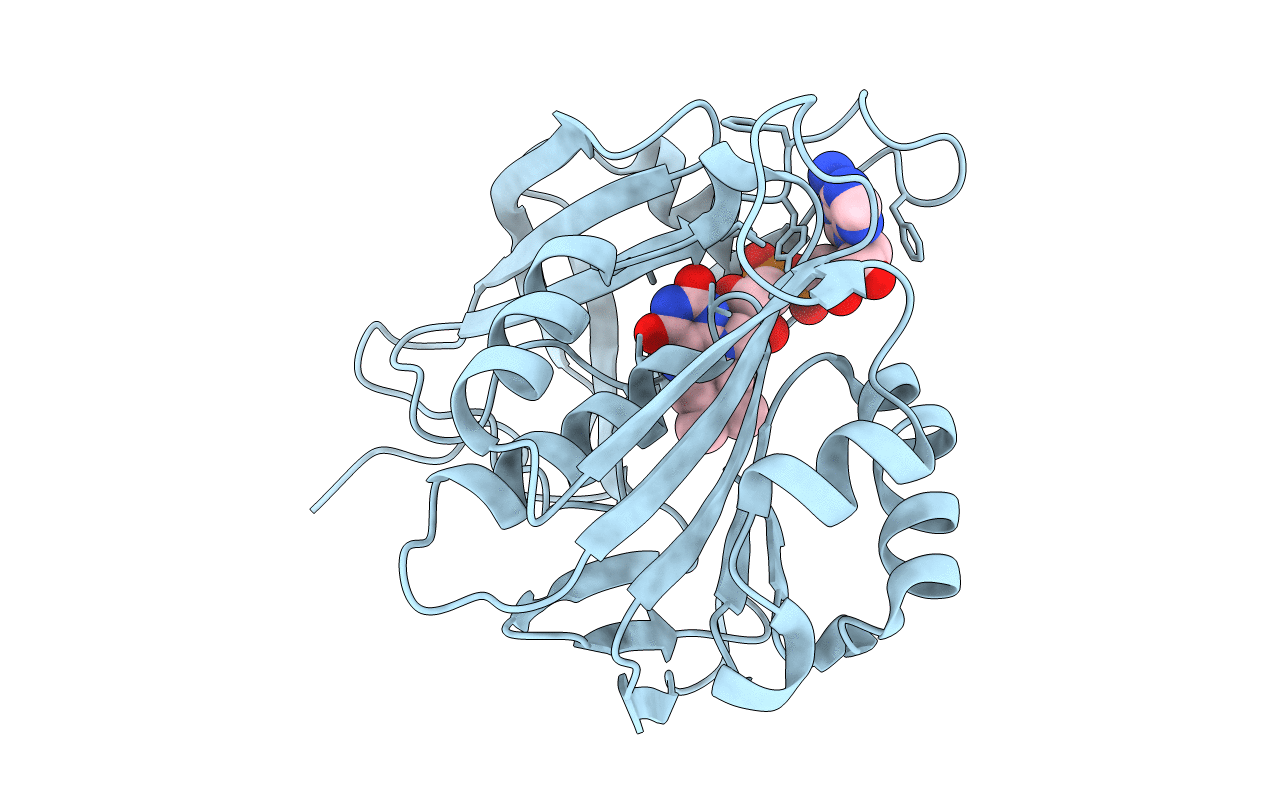
Title:
The Structure of Human Erythrocyte NADH-cytochrome b5 Reductase
Biological Source:
Source Organism(s):
Homo sapiens (Taxon ID: 9606)
Expression System(s):
Method Details:
Experimental Method:
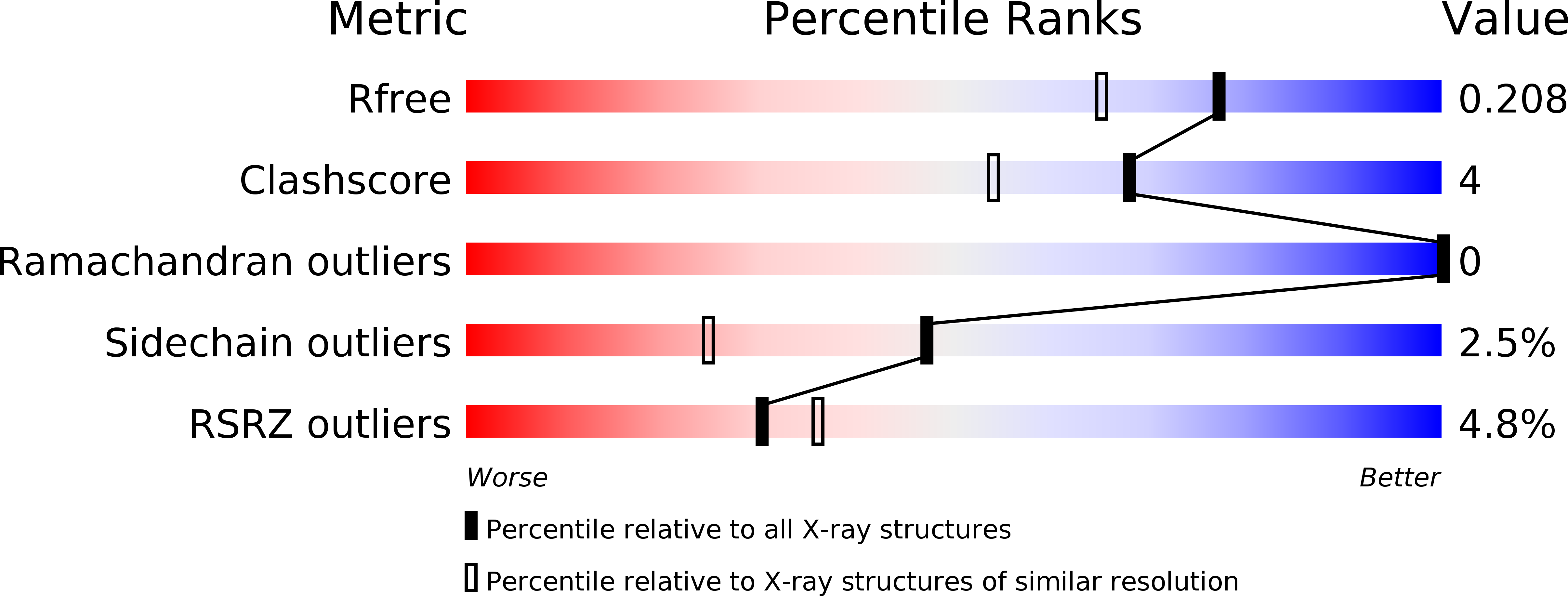
Resolution:
1.75 Å
R-Value Free:
0.20
R-Value Work:
0.16
R-Value Observed:
0.16
Space Group:
P 41