1UMJ image
Deposition Date 2003-10-02
Release Date 2004-10-05
Last Version Date 2023-10-25
Entry Detail
PDB ID:
1UMJ
Title:
Crystal structure of Pyrococcus horikoshii CutA in the presence of 3M guanidine hydrochloride
Biological Source:
Source Organism:
Method Details:
Experimental Method:
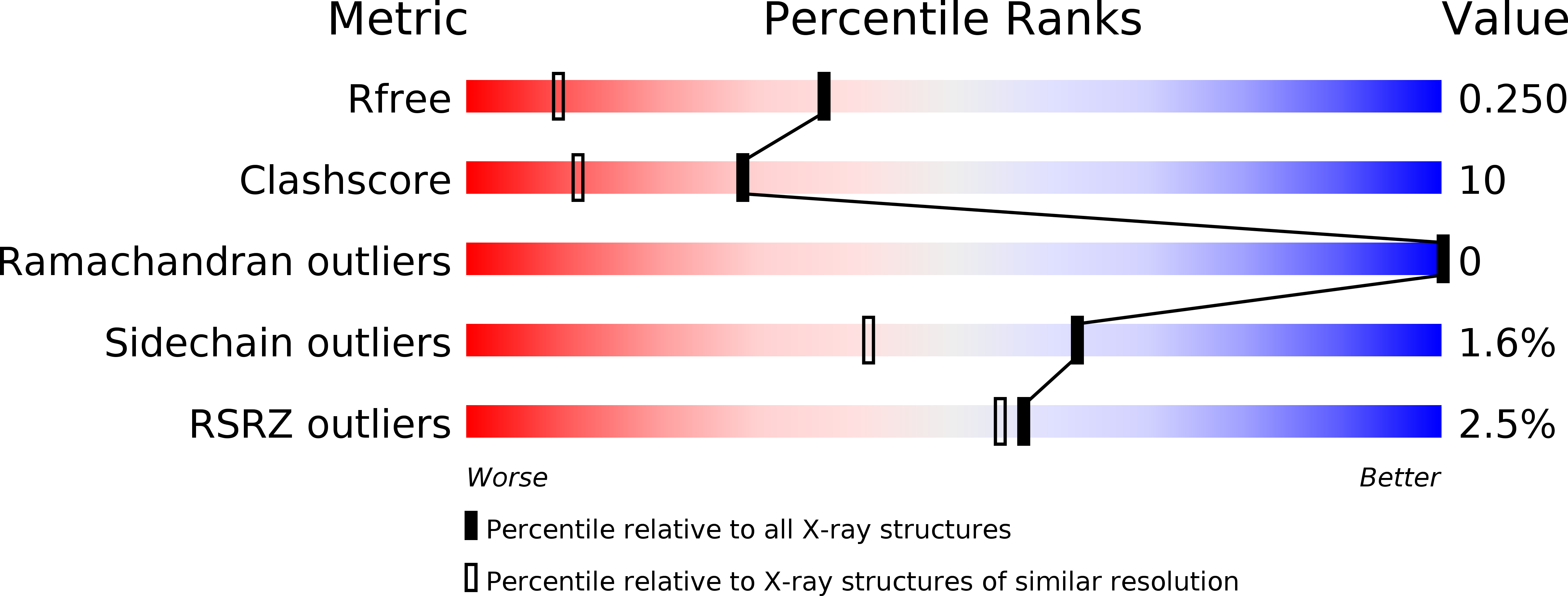
Resolution:
1.60 Å
R-Value Free:
0.25
R-Value Work:
0.21
Space Group:
P 3
Macromolecular Entities
Structures with similar UniProt ID
Protein Blast
Polymer Type:polypeptide(L)
Molecule:periplasmic divalent cation tolerance protein CutA
Gene (Uniprot):cutA
Chain IDs:A, B
Chain Length:102
Number of Molecules:2
Biological Source:Pyrococcus horikoshii
Ligand Molecules
Primary Citation
Structural evidence for guanidine-protein side chain interactions: crystal structure of CutA from Pyrococcus horikoshii in 3M guanidine hydrochloride
Biochem.Biophys.Res.Commun. 323 185 191 (2004)
PMID: 15351719 DOI: 10.1016/j.bbrc.2004.08.081

Abstact

This study was carried out to investigate the structural perturbation of the protein's local structure by the denaturants under non-denaturing conditions. Crystal structure of CutA from an archaeon Pyrococcus horikosii (PhoCutA), a heavy-metal binding protein, was determined at 1.6-angstroms resolution in the presence of 3 M guanidine HCl (GdnHCl). Native PhoCutA has a large number of short intramolecular hydrogen bonds and salt bridges on the protein surface, of which greater than 90% of hydrogen bonds and all salt bridges were retained in 3 M GdnHCl. Hydrogen bonds that disappeared in the GdnHCl crystal structure were mainly located on the protein surface, especially around the structurally perturbed loop, suggesting interactions between peptide groups and GdnHCl. Only a few GdnH+ ions were observed in the crystal structure, although none at the surface, of the protein. Two GdnH+ ions were observed in the center of the trimeric structure, replacing water molecules, and were hydrogen bonded with Asp84 and Asp86 of each chain. The exterior loop from Tyr39 to Lys44, including Trp40-Trp41, was perturbed structurally. Decreases in temperature factors were observed in beta strand 5 and the N terminus of helix 3. These results suggest the specific bindings of GdnH+ with some acidic residues and the non-specific bindings around Trp residues and peptide groups on the protein surface and that binding of GdnHCl to the native protein is limited, resulting in local structural perturbation.

Legend

Protein

Chemical

Disease

Primary Citation of related structures
Feedback Form
Name
Email
Institute
Feedback