1ULZ image
Deposition Date 2003-09-18
Release Date 2004-03-09
Last Version Date 2023-12-27
Entry Detail
PDB ID:
1ULZ
Keywords:
Title:
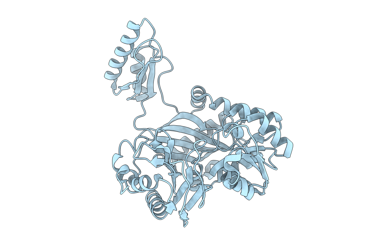
Crystal structure of the biotin carboxylase subunit of pyruvate carboxylase
Biological Source:
Source Organism:
Host Organism:
Method Details:
Experimental Method:
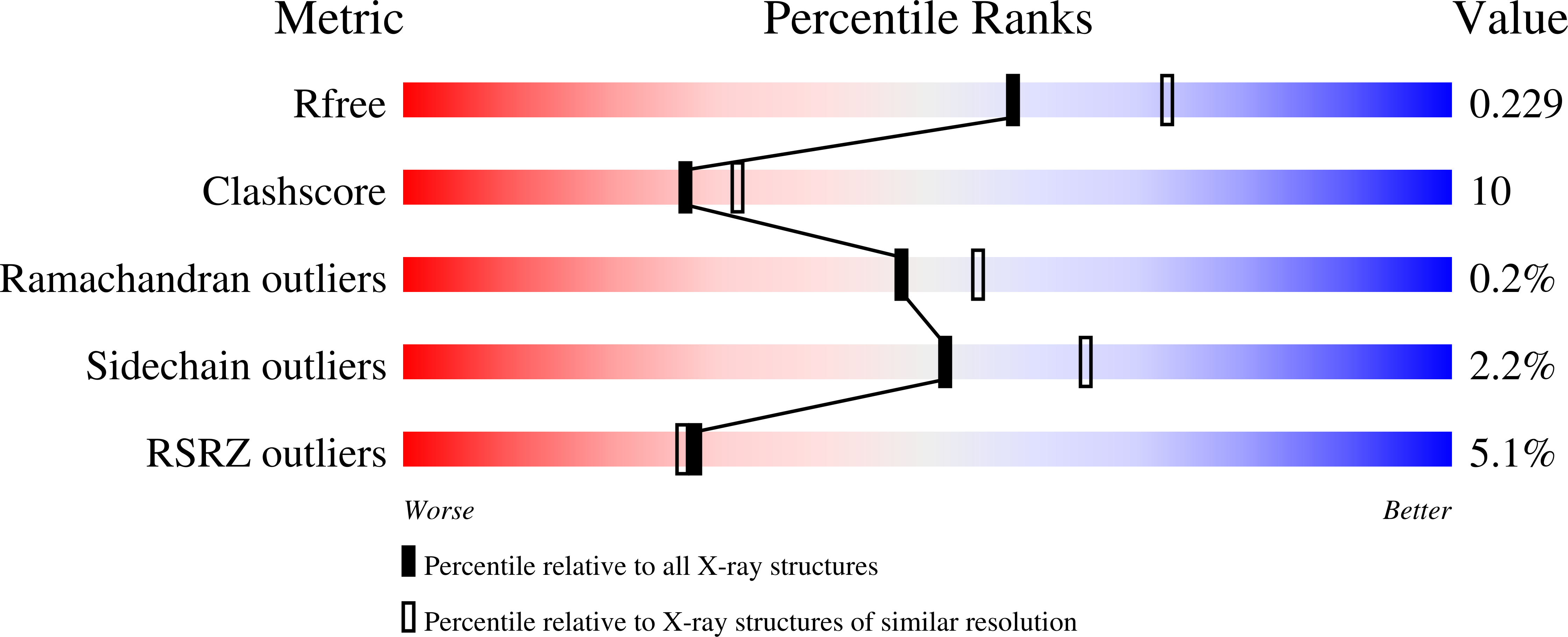
Resolution:
2.20 Å
R-Value Free:
0.23
R-Value Work:
0.19
R-Value Observed:
0.20
Space Group:
P 21 21 2
Macromolecular Entities
Structures with similar UniProt ID
Protein Blast
Polymer Type:polypeptide(L)
Molecule:pyruvate carboxylase n-terminal domain
Gene (Uniprot):pycB
Mutagens:F2V
Chain IDs:A
Chain Length:451
Number of Molecules:1
Biological Source:Aquifex aeolicus
Primary Citation
Structure of the biotin carboxylase subunit of pyruvate carboxylase from Aquifex aeolicus at 2.2 A resolution.
Acta Crystallogr.,Sect.D 60 486 492 (2004)
PMID: 14993673 DOI: 10.1107/S0907444904000423

Abstact

Pyruvate carboxylase (PC) is distributed in many eukaryotes as well as in some prokaryotes. PC catalyzes the ATP-dependent carboxylation of pyruvate to form oxalacetate. PC has three functional domains, one of which is a biotin carboxylase (BC) domain. The BC subunit of PC from Aquifex aeolicus (PC-beta) was crystallized in an orthorhombic form with space group P2(1)2(1)2, unit-cell parameters a = 92.4, b = 122.1, c = 59.0 A and one molecule in the asymmetric unit. Diffraction data were collected at 100 K on BL24XU at SPring-8. The crystal structure was determined by the molecular-replacement method and refined against 20.0-2.2 A resolution data, giving an R factor of 0.199 and a free R factor of 0.236. The crystal structure revealed that PC-beta forms a dimeric quaternary structure consisting of two molecules related by crystallographic twofold symmetry. The overall structure of PC-beta is similar to other biotin-dependent carboxylases, such as acetyl-CoA carboxylase (ACC). Although some parts of domain B were disordered in ACC, the corresponding parts of PC-beta were clearly determined in the crystal structure. From comparison between the active-site structure of ACC with ATP bound and a virtual model of PC-beta with ATP bound, it was shown that the backbone torsion angles of Glu203 in PC-beta change and some of water molecules in the active site of PC-beta are excluded upon ATP binding.

Legend

Protein

Chemical

Disease

Primary Citation of related structures