1UL3 image
Deposition Date 2003-09-09
Release Date 2003-12-16
Last Version Date 2023-10-25
Entry Detail
PDB ID:
1UL3
Title:
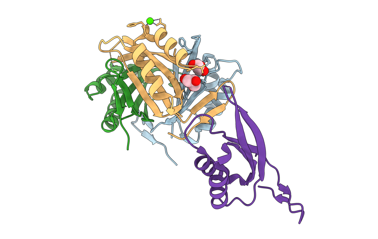
Crystal Structure of PII from Synechocystis sp. PCC 6803
Biological Source:
Source Organism:
Host Organism:
Method Details:
Experimental Method:
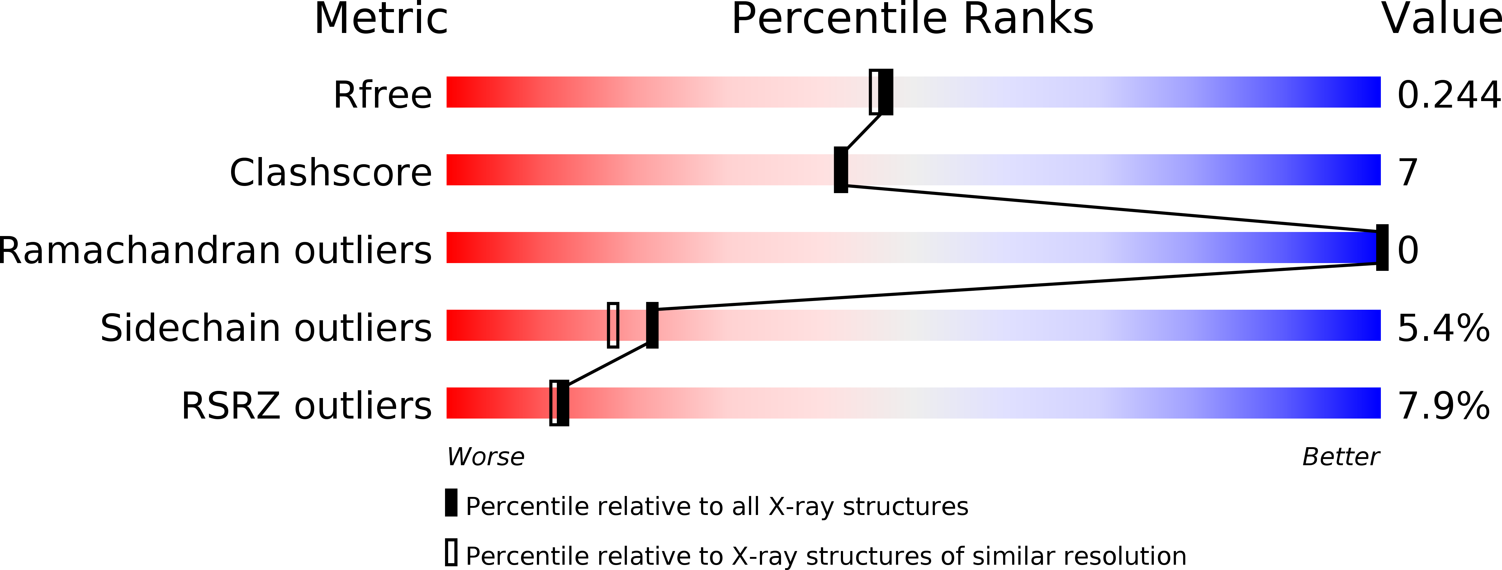
Resolution:
2.00 Å
R-Value Free:
0.25
R-Value Work:
0.21
R-Value Observed:
0.22
Space Group:
H 3
Macromolecular Entities
Structures with similar UniProt ID
Protein Blast
Polymer Type:polypeptide(L)
Molecule:Nitrogen regulatory protein P-II
Gene (Uniprot):glnB
Chain IDs:A, B, C, D
Chain Length:112
Number of Molecules:4
Biological Source:Synechocystis sp.
Primary Citation
The structures of the PII proteins from the cyanobacteria Synechococcus sp. PCC 7942 and Synechocystis sp. PCC 6803.
Acta Crystallogr.,Sect.D 59 2183 2190 (2003)
PMID: 14646076 DOI: 10.1107/S0907444903019589

Abstact

The PII proteins from the cyanobacteria Synechococcus sp. PCC 7942 and Synechocystis sp. PCC 6803 have been crystallized and high-resolution structures have been obtained using X-ray crystallography. The core of these new structures is similar to that of the PII proteins from Escherichia coli, although the structures of the T- and C-loops differ. The T-loop of the Synechococcus protein is ordered, but appears to be stabilized by crystal contacts. The same loop in the Synechocystis protein is disordered. The C-terminus of the Synechocystis protein is stabilized by hydrogen bonding to the same region of a crystallographically related molecule. The same terminus in the Synechococcus protein is stabilized by coordination with a metal ion. These observations are consistent with the idea that both the T-loop and the C-terminus of PII proteins are flexible in solution and that this flexibility may be important for receptor recognition. Sequence comparisons are used to identify regions of the sequence unique to the cyanobacteria.

Legend

Protein

Chemical

Disease

Primary Citation of related structures