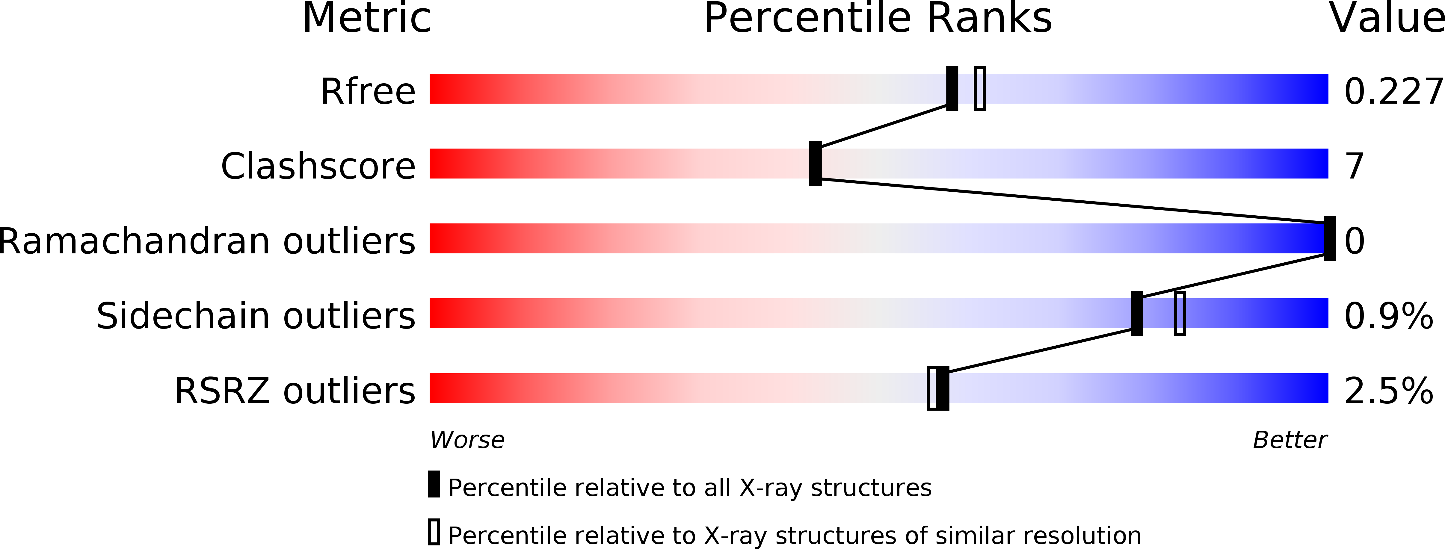
1UII image
Deposition Date 2003-07-16
Release Date 2004-07-16
Last Version Date 2023-12-27
Entry Detail
PDB ID:
1UII
Keywords:
Title:
Crystal structure of Geminin coiled-coil domain
Biological Source:
Source Organism:
Homo sapiens (Taxon ID: 9606)
Host Organism:
Method Details:
Experimental Method:
Resolution:
2.00 Å
R-Value Free:
0.23
R-Value Work:
0.21
R-Value Observed:
0.21
Space Group:
P 21 21 21
Macromolecular Entities
Polymer Type:polypeptide(L)
Molecule:Geminin
Gene (Uniprot):GMNN
Chain IDs:A, B
Chain Length:83
Number of Molecules:2
Biological Source:Homo sapiens
Primary Citation
A dimerized coiled-coil domain and an adjoining part of geminin interact with two sites on Cdt1 for replication inhibition
Mol.Cell 15 245 258 (2004)
PMID: 15260975 DOI: 10.1016/j.molcel.2004.06.045

Abstact

Geminin is a cellular protein that associates with Cdt1 and inhibits Mcm2-7 loading during S phase. It prevents multiple cycles of replication per cell cycle and prevents episome replication. It also directly inhibits the HoxA11 transcription factor. Here we report that geminin forms a parallel coiled-coil homodimer with atypical residues in the dimer interface. Point mutations that disrupt the dimerization abolish interaction with Cdt1 and inhibition of replication. An array of glutamic acid residues on the coiled-coil domain surface interacts with positive charges in the middle of Cdt1. An adjoining region interacts independently with the N-terminal 100 residues of Cdt1. Both interactions are essential for replication inhibition. The negative residues on the coiled-coil domain and a different part of geminin are also required for interaction with HoxA11. Therefore a rigid cylinder with negative surface charges is a critical component of a bipartite interaction interface between geminin and its cellular targets.

Legend

Protein

Chemical

Disease

Primary Citation of related structures