
Deposition Date
1996-11-26
Release Date
1997-11-26
Last Version Date
2024-10-16
Entry Detail
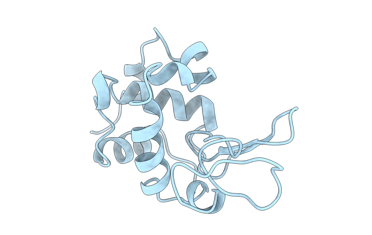
PDB ID:
1UIG
Keywords:
Title:
ANALYSIS OF THE STABILIZATION OF HEN LYSOZYME WITH THE HELIX DIPOLE AND CHARGED SIDE CHAINS
Biological Source:
Source Organism(s):
Gallus gallus (Taxon ID: 9031)
Expression System(s):
Method Details:
Experimental Method:
Resolution:
1.95 Å
R-Value Work:
0.16
R-Value Observed:
0.16
Space Group:
P 43 21 2
