1UHB image
Deposition Date 2003-06-27
Release Date 2004-07-27
Last Version Date 2024-11-13
Entry Detail
PDB ID:
1UHB
Keywords:
Title:
Crystal structure of porcine alpha trypsin bound with auto catalyticaly produced native peptide at 2.15 A resolution
Biological Source:
Source Organism:
Sus scrofa (Taxon ID: 9823)
Method Details:
Experimental Method:
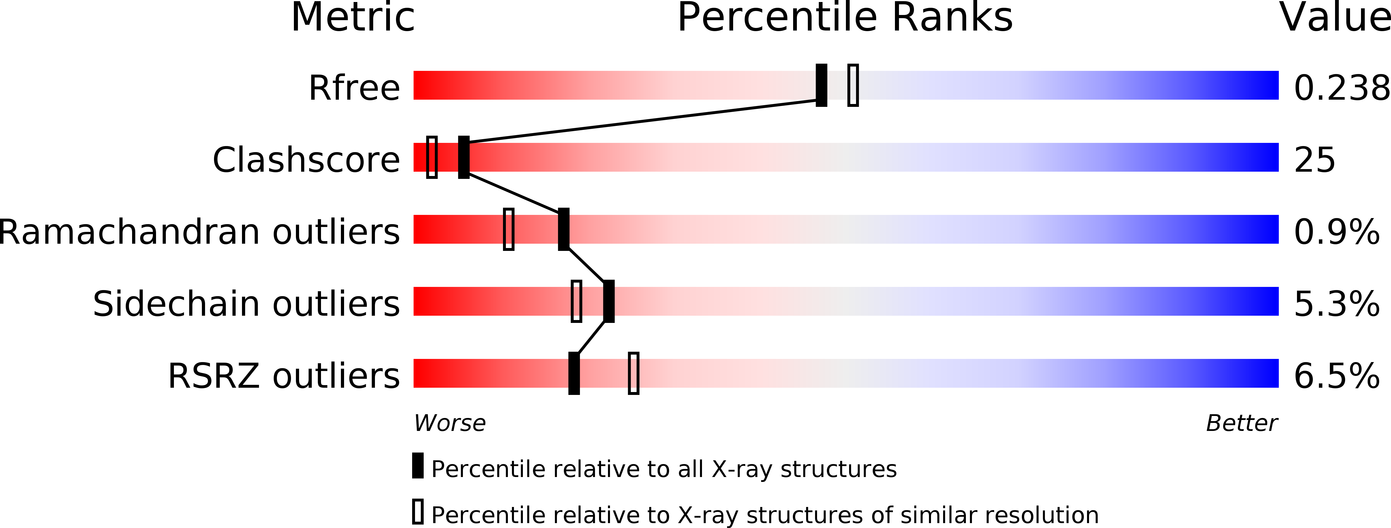
Resolution:
2.15 Å
R-Value Free:
0.22
R-Value Work:
0.19
R-Value Observed:
0.19
Space Group:
P 21 21 21
Macromolecular Entities
Structures with similar UniProt ID
Protein Blast
Polymer Type:polypeptide(L)
Molecule:Trypsin
Chain IDs:A
Chain Length:125
Number of Molecules:1
Biological Source:Sus scrofa
Structures with similar UniProt ID
Protein Blast
Polymer Type:polypeptide(L)
Molecule:Trypsin
Chain IDs:B
Chain Length:98
Number of Molecules:1
Biological Source:Sus scrofa
Structures with similar UniProt ID
Protein Blast
Polymer Type:polypeptide(L)
Molecule:9-mer peptide from Trypsin
Chain IDs:C (auth: P)
Chain Length:9
Number of Molecules:1
Biological Source:Sus scrofa
Primary Citation
Trypsin activity reduced by an autocatalytically produced nonapeptide.
J.Biomol.Struct.Dyn. 21 737 744 (2004)
PMID: 15106996 DOI: 10.1080/07391102.2004.10506964

Abstact

Trypsin, a serine protease enzyme plays a pivotal role in digestion and is autocatalytic. The crystal structure of a complex formed between porcine trypsin and an auto catalytically produced peptide is reported here. This complex shows a reduction in enzyme activity as compared to native beta-trypsin. The nonapeptide has a lysine, which is recognized by Asp 189 at the specificity pocket. The auto catalytically produced native nonapeptide is bound at the active site cleft like other trypsin inhibitors but the important interactions with the oxyanion hole are absent. The peptide covers only a part of the active site cleft and hence the enzyme activity is reduced rather than being inhibited.

Legend

Protein

Chemical

Disease

Primary Citation of related structures