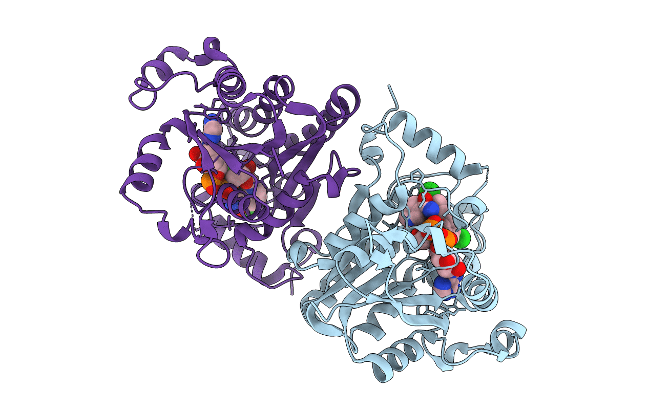
1UH5 image
Deposition Date 2003-06-24
Release Date 2004-09-28
Last Version Date 2023-10-25
Entry Detail
PDB ID:
1UH5
Keywords:
Title:
Crystal Structure of Enoyl-ACP Reductase with Triclosan at 2.2angstroms
Biological Source:
Source Organism:
Method Details:
Experimental Method:
Resolution:
2.20 Å
R-Value Free:
0.21
R-Value Work:
0.18
R-Value Observed:
0.18
Space Group:
P 43 21 2
Macromolecular Entities
Polymer Type:polypeptide(L)
Molecule:enoyl-ACP reductase
Chain IDs:A, B
Chain Length:329
Number of Molecules:2
Biological Source:Plasmodium falciparum
Primary Citation
Structural basis for the variation in triclosan affinity to enoyl reductases.
J.Mol.Biol. 343 147 155 (2004)
PMID: 15381426 DOI: 10.1016/j.jmb.2004.08.033

Abstact

Bacteria synthesize fatty acids in a dissociated type pathway different from that in humans. Enoyl acyl carrier protein reductase, which catalyzes the final step of fatty acid elongation, has been validated as a potential anti-microbial drug target. Triclosan is known to inhibit this enzyme effectively. Precise characterization of the mode of triclosan binding is required to develop highly specific inhibitors. With this in view, interactions between triclosan, the cofactor NADH/NAD+ and the enzyme from five different species, one plant and four of microbial origin, have been examined in the available crystal structures. A comparison of these structures shows major structural differences at the substrate/inhibitor/cofactor-binding loop. The analysis reveals that the conformation of this flexible loop and the binding affinities of triclosan to each of these enzymes are strongly correlated.

Legend

Protein

Chemical

Disease

Primary Citation of related structures