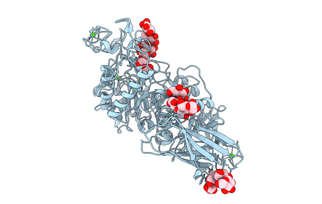
1UH2 image
Deposition Date 2003-06-23
Release Date 2004-01-13
Last Version Date 2023-12-27
Entry Detail
PDB ID:
1UH2
Keywords:
Title:
Thermoactinomyces vulgaris R-47 alpha-amylase/malto-hexaose complex
Biological Source:
Source Organism:
Host Organism:
Method Details:
Experimental Method:
Resolution:
2.00 Å
R-Value Free:
0.21
R-Value Work:
0.18
R-Value Observed:
0.18
Space Group:
C 1 2 1
Macromolecular Entities
Structures with similar UniProt ID
Protein Blast
Polymer Type:polypeptide(L)
Molecule:alpha-amylase I
Mutagens:D356N, E396Q
Chain IDs:A
Chain Length:637
Number of Molecules:1
Biological Source:Thermoactinomyces vulgaris
Ligand Molecules
Peptide-like Molecules
PRD_900010
PRD_900030
Primary Citation
Complex Structures of Thermoactinomyces vulgaris R-47 alpha-Amylase 1 with Malto-oligosaccharides Demonstrate the Role of Domain N Acting as a Starch-binding Domain
J.Mol.Biol. 335 811 822 (2004)
PMID: 14687576 DOI: 10.1016/j.jmb.2003.10.078

Abstact

The X-ray structures of complexes of Thermoactinomyces vulgaris R-47 alpha-amylase 1 (TVAI) with an inhibitor acarbose and an inactive mutant TVAI with malto-hexaose and malto-tridecaose have been determined at 2.6, 2.0 and 1.8A resolution, and the structures have been refined to R-factors of 0.185 (R(free)=0.225), 0.184 (0.217) and 0.164 (0.200), respectively, with good chemical geometries. Acarbose binds to the catalytic site of TVAI, and interactions between acarbose and the enzyme are very similar to those found in other structure-solved alpha-amylase/acarbose complexes, supporting the proposed catalytic mechanism. Based on the structure of the TVAI/acarbose complex, the binding mode of pullulan containing alpha-(1,6) glucoside linkages could be deduced. Due to the structural difference caused by the replaced amino acid residue (Gln396 for Glu) in the catalytic site, malto-hexaose and malto-tridecaose partially bind to the catalytic site, giving a mimic of the enzyme/product complex. Besides the catalytic site, four sugar-binding sites on the molecular surface are found in these X-ray structures. Two sugar-binding sites in domain N hold the oligosaccharides with a regular helical structure of amylose, which suggests that the domain N is a starch-binding domain acting as an anchor to starch in the catalytic reaction of the enzyme. An assay of hydrolyzing activity for the raw starches confirmed that TVAI can efficiently hydrolyze raw starch.

Legend

Protein

Chemical

Disease

Primary Citation of related structures
Feedback Form
Name
Email
Institute
Feedback