1UFI image
Deposition Date 2003-05-30
Release Date 2004-02-17
Last Version Date 2023-12-27
Entry Detail
PDB ID:
1UFI
Title:
Crystal structure of the dimerization domain of human CENP-B
Biological Source:
Source Organism:
Homo sapiens (Taxon ID: 9606)
Method Details:
Experimental Method:
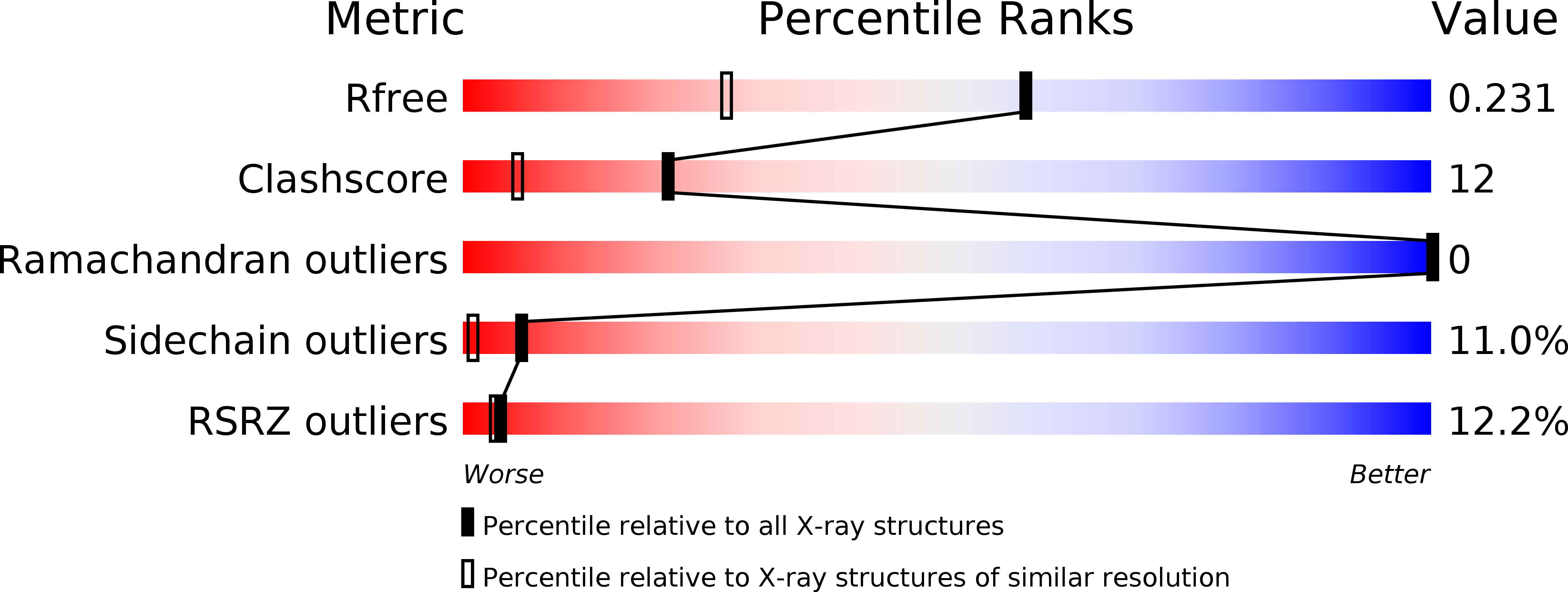
Resolution:
1.65 Å
R-Value Free:
0.30
R-Value Work:
0.23
R-Value Observed:
0.23
Space Group:
P 21 21 21
Macromolecular Entities
Polymer Type:polypeptide(L)
Molecule:Major centromere autoantigen B
Gene (Uniprot):CENPB
Chain IDs:A, B, C, D
Chain Length:64
Number of Molecules:4
Biological Source:Homo sapiens
Primary Citation
Crystal structure of the human centromere protein B (CENP-B) dimerization domain at 1.65-A resolution
J.Biol.Chem. 278 51454 51461 (2003)
PMID: 14522975 DOI: 10.1074/jbc.M310388200

Abstact

The human centromere protein B (CENP-B), a centromeric heterochromatin component, forms a homodimer that specifically binds to a distinct DNA sequence (the CENP-B box), which appears within every other alpha-satellite repeat. Previously, we determined the structure of the human CENP-B DNA-binding domain, CENP-B-(1-129), complexed with the CENP-B box DNA. In the present study, we determined the crystal structure of its dimerization domain (CENP-B-(540-599)), another functional domain of CENP-B, at 1.65-A resolution. CENP-B-(540-599) contains two alpha-helices, which are folded into an antiparallel configuration. The CENP-B-(540-599) dimer formed a symmetrical, antiparallel, four-helix bundle structure with a large hydrophobic patch in which 23 residues of one monomer form van der Waals contacts with the other monomer. In the CENP-B-(540-599) dimer, the N-terminal ends of CENP-B-(540-599) are oriented on opposite sides of the dimer. This CENP-B dimer configuration may be suitable for capturing two distant CENP-B boxes during centromeric heterochromatin formation.

Legend

Protein

Chemical

Disease

Primary Citation of related structures