1UED image
Deposition Date 2003-05-11
Release Date 2003-12-09
Last Version Date 2023-10-25
Entry Detail
PDB ID:
1UED
Keywords:
Title:
Crystal Structure of OxyC a Cytochrome P450 Implicated in an Oxidative C-C Coupling Reaction During Vancomycin Biosynthesis.
Biological Source:
Source Organism:
Host Organism:
Method Details:
Experimental Method:
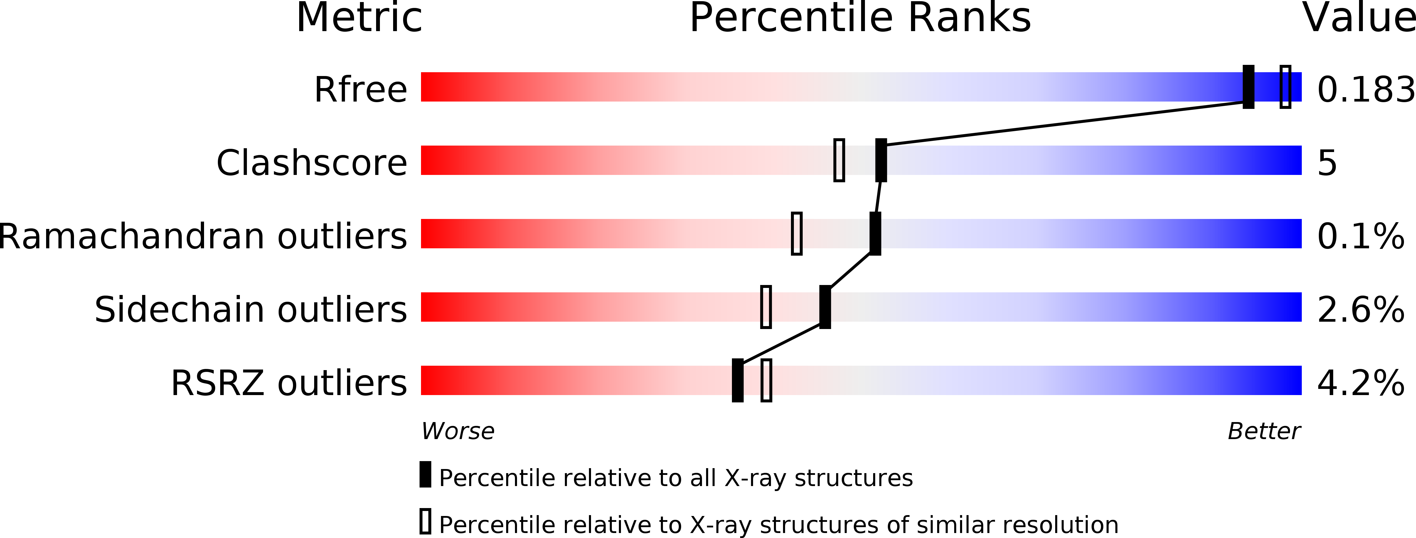
Resolution:
1.90 Å
R-Value Free:
0.23
R-Value Work:
0.19
R-Value Observed:
0.19
Space Group:
P 1 21 1
Macromolecular Entities
Structures with similar UniProt ID
Protein Blast
Polymer Type:polypeptide(L)
Molecule:P450 monooxygenase
Gene (Uniprot):cyp165C4
Chain IDs:A, B
Chain Length:406
Number of Molecules:2
Biological Source:Amycolatopsis orientalis
Primary Citation
Crystal structure of OxyC, a cytochrome P450 implicated in an oxidative C-C coupling reaction during vancomycin biosynthesis
J.Biol.Chem. 278 46727 46733 (2003)
PMID: 12888556 DOI: 10.1074/jbc.M306486200

Abstact

Gene inactivation studies point to the involvement of OxyC in catalyzing the last oxidative phenol coupling reaction during glycopeptide antibiotic biosynthesis. Presently, the substrate and exact timing of the OxyC reaction are unknown. The substrate might be the bicyclic heptapeptide or a thioester derivative bound to a protein carrier domain. OxyC from the vancomycin producer Amycolatopsis orientalis was produced in Escherichia coli and crystallized, and its structure was determined to 1.9 A resolution. OxyC gave UV-visible spectra characteristic of a P450-like hemoprotein in the low spin ferric state. After reduction to the ferrous state by dithionite the CO-ligated form gave a 450-nm peak in a UV-difference spectrum. The addition of vancomycin aglycone to OxyC produced type I changes to the UV spectrum. OxyC exhibits the typical P450-fold, with the Cys ligand loop containing the signature sequence FGHGX-HXCLG and Cys-356 being the proximal axial thiolate ligand of the heme iron. The observation of a water molecule bound to the heme iron is consistent with the UV-visible spectra of OxyC indicating a low spin heme. A polyethylene glycol molecule occupying the active site might mimic the bicyclic heptapeptide substrate. Analysis of the structure of Oxy-proteins and other P450s indicates regions that might be involved in binding of the redox partner and possibly the protein carrier domain.

Legend

Protein

Chemical

Disease

Primary Citation of related structures