1UE8 image
Deposition Date 2003-05-09
Release Date 2004-07-13
Last Version Date 2023-12-27
Entry Detail
PDB ID:
1UE8
Title:
Crystal Structure of Thermophilic Cytochrome P450 from Sulfolobus tokodaii
Biological Source:
Source Organism:
Host Organism:
Method Details:
Experimental Method:
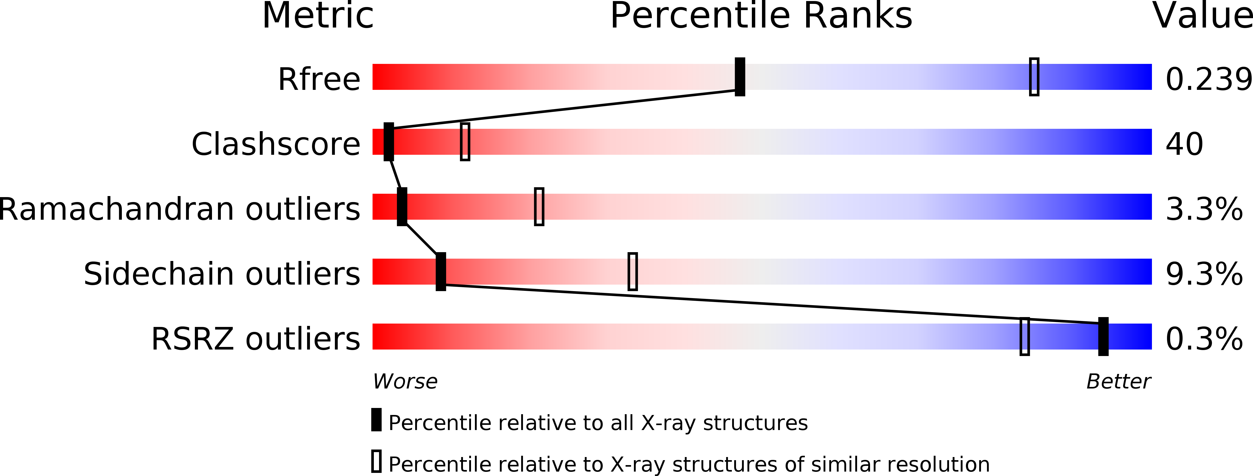
Resolution:
3.00 Å
R-Value Free:
0.23
R-Value Work:
0.19
R-Value Observed:
0.19
Space Group:
P 21 21 21
Macromolecular Entities
Structures with similar UniProt ID
Protein Blast
Polymer Type:polypeptide(L)
Molecule:367aa long hypothetical cytochrome P450
Gene (Uniprot):cyp119
Chain IDs:A
Chain Length:367
Number of Molecules:1
Biological Source:Sulfolobus tokodaii
Primary Citation
Structure and direct electrochemistry of cytochrome P450 from the thermoacidophilic crenarchaeon, Sulfolobus tokodaii strain 7
J.Inorg.Biochem. 98 1194 1199 (2004)
PMID: 15219985 DOI: 10.1016/j.jinorgbio.2004.05.002

Abstact

Cytochrome P450 from thermoacidophilic crenarchaeon, Sulfolobus tokodaii strain 7 (P450st) has been expressed in Escherichia coli and purified at high homogeneity. P450st was crystallized in an orthorhombic system with the space group P2(1)2(1)2(1) and cell dimensions of a=53.6 A, b=55.1 A, and c=130.9 A, and the structure was determined at a 3.0 A resolution. The final R-factor was 0.194 (Rfree=0.235). Structural comparison with cytochrome P450 from S. solfataricus (CYP119) suggests that the region composed of the F to G helices and the Cl- binding site is responsible for the affinity for a ligand coordinating heme iron. Direct electrochemistry of P450st in a didodecyldimethylammonium bromide (DDAB) film on a plastic formed carbon (PFC) electrode has also been demonstrated. A quasi-reversible redox response has been observed even at elevated temperatures of up to 80 degrees C.

Legend

Protein

Chemical

Disease

Primary Citation of related structures