Deposition Date
1995-10-30
Release Date
1996-03-08
Last Version Date
2024-02-14
Entry Detail
PDB ID:
1UDI
Keywords:
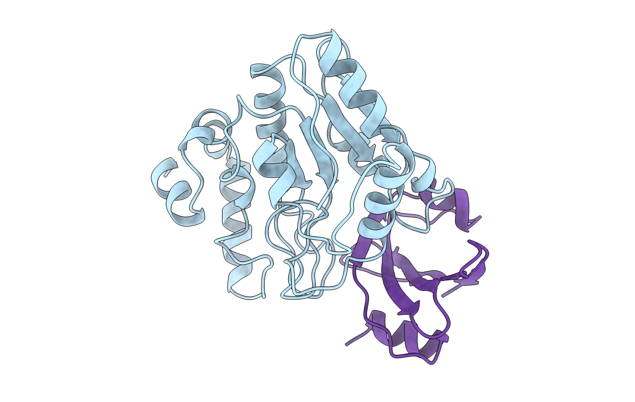
Title:
NUCLEOTIDE MIMICRY IN THE CRYSTAL STRUCTURE OF THE URACIL-DNA GLYCOSYLASE-URACIL GLYCOSYLASE INHIBITOR PROTEIN COMPLEX
Biological Source:
Source Organism(s):
Herpes simplex virus (type 1 / strain 17) (Taxon ID: 10299)
Bacillus phage PBS1 (Taxon ID: 10683)
Bacillus phage PBS1 (Taxon ID: 10683)
Method Details:
Experimental Method:
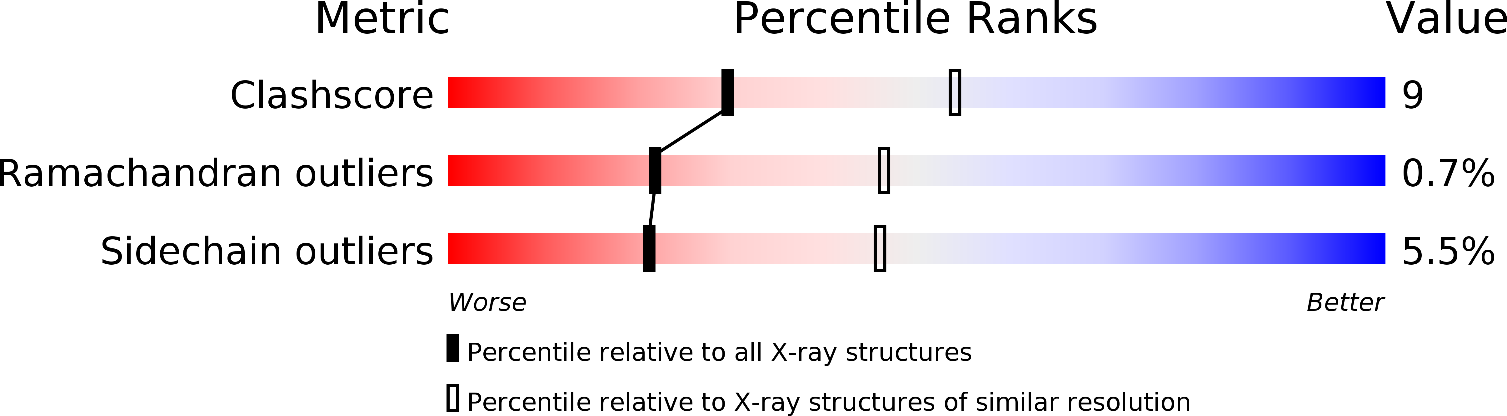
Resolution:
2.70 Å
R-Value Free:
0.27
R-Value Work:
0.18
R-Value Observed:
0.18
Space Group:
P 65