Deposition Date
2003-04-28
Release Date
2003-07-22
Last Version Date
2023-10-25
Entry Detail
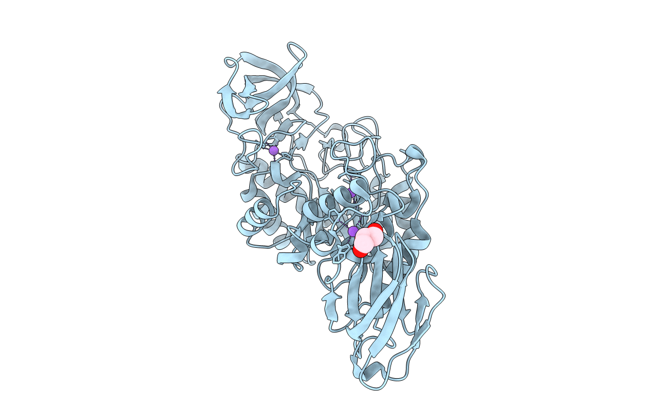
PDB ID:
1UD2
Keywords:
Title:
Crystal structure of calcium-free alpha-amylase from Bacillus sp. strain KSM-K38 (AmyK38)
Biological Source:
Source Organism(s):
Bacillus sp. KSM-K38 (Taxon ID: 129736)
Expression System(s):
Method Details:
Experimental Method:
Resolution:
2.13 Å
R-Value Free:
0.22
R-Value Work:
0.19
Space Group:
P 2 3