
Deposition Date
2003-04-04
Release Date
2003-04-29
Last Version Date
2024-11-13
Entry Detail
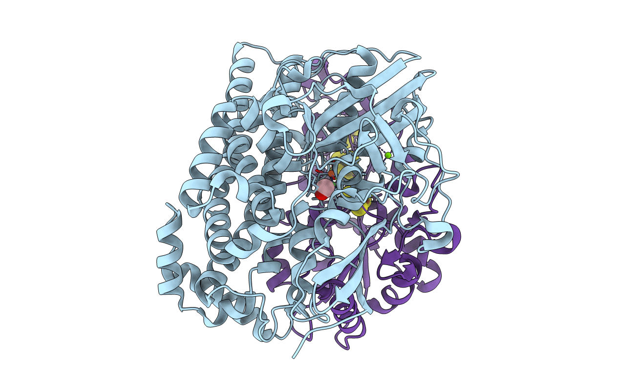
PDB ID:
1UBJ
Keywords:
Title:
Three-dimensional Structure of The Carbon Monoxide Complex of [NiFe]hydrogenase From Desulufovibrio vulgaris Miyazaki F
Biological Source:
Source Organism(s):
Desulfovibrio vulgaris str. 'Miyazaki F' (Taxon ID: 883)
Method Details:
Experimental Method:
Resolution:
1.35 Å
R-Value Free:
0.18
R-Value Work:
0.12
R-Value Observed:
0.12
Space Group:
P 21 21 21
