1UBC image
Entry Detail
PDB ID:
1UBC
Keywords:
Title:
Structure of Reca Protein
Biological Source:
Source Organism:
Host Organism:
PDB Version:
Deposition Date:
2003-04-04
Release Date:
2003-07-22
Method Details:
Experimental Method:
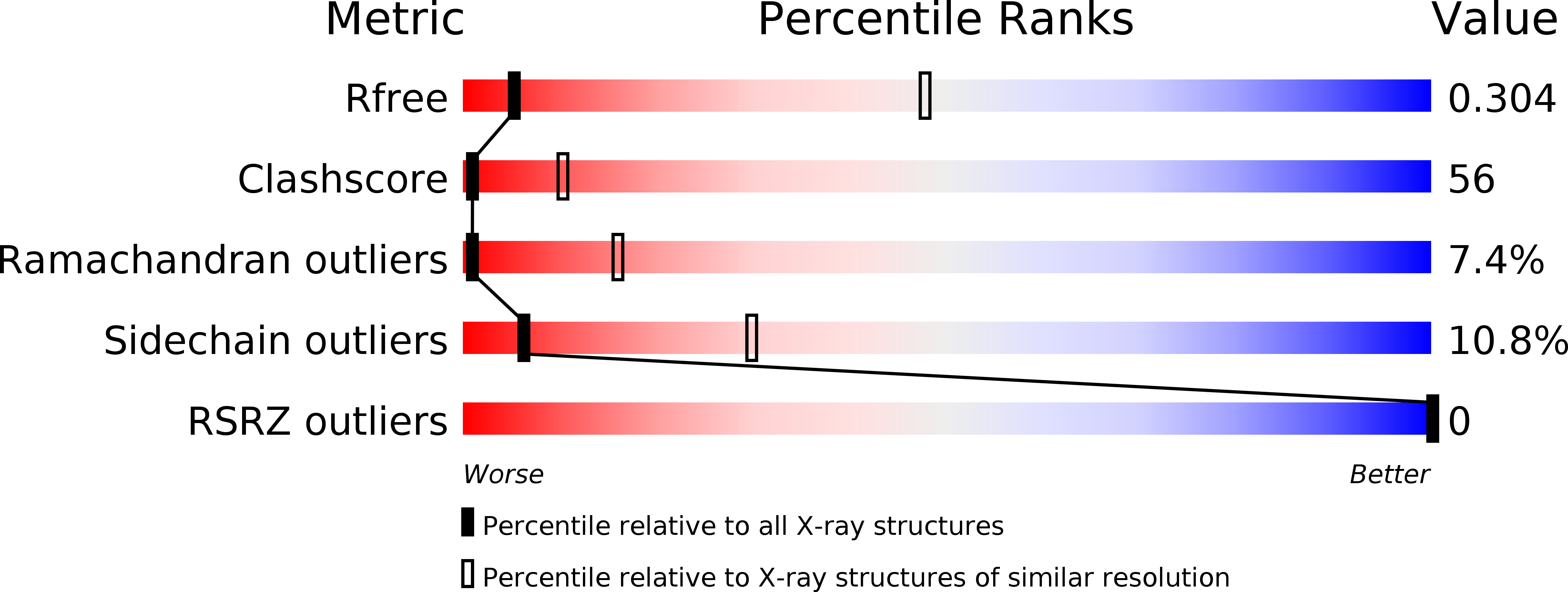
Resolution:
3.80 Å
R-Value Free:
0.30
R-Value Work:
0.24
R-Value Observed:
0.24
Space Group:
P 61
Macromolecular Entities
Polymer Type:polypeptide(L)
Description:RecA
Chain IDs:A
Chain Length:349
Number of Molecules:1
Biological Source:Mycobacterium smegmatis
Primary Citation
Crystal Structures of Mycobacterium smegmatis RecA and Its Nucleotide Complexes
J.BACTERIOL. 185 4280 4284 (2003)
PMID: 12837805 DOI: 10.1128/JB.185.14.4280-4284.2003

Abstact

The crystal structures of Mycobacterium smegmatis RecA (RecA(Ms)) and its complexes with ADP, ATPgammaS, and dATP show that RecA(Ms) has an expanded binding site like that in Mycobacterium tuberculosis RecA, although there are small differences between the proteins in their modes of nucleotide binding. Nucleotide binding is invariably accompanied by the movement of Gln 196, which appears to provide the trigger for transmitting the effect of nucleotide binding to the DNA-binding loops. These observations provide a framework for exploring the known properties of the RecA proteins.

Legend

Protein

Chemical

Disease

Primary Citation of related structures