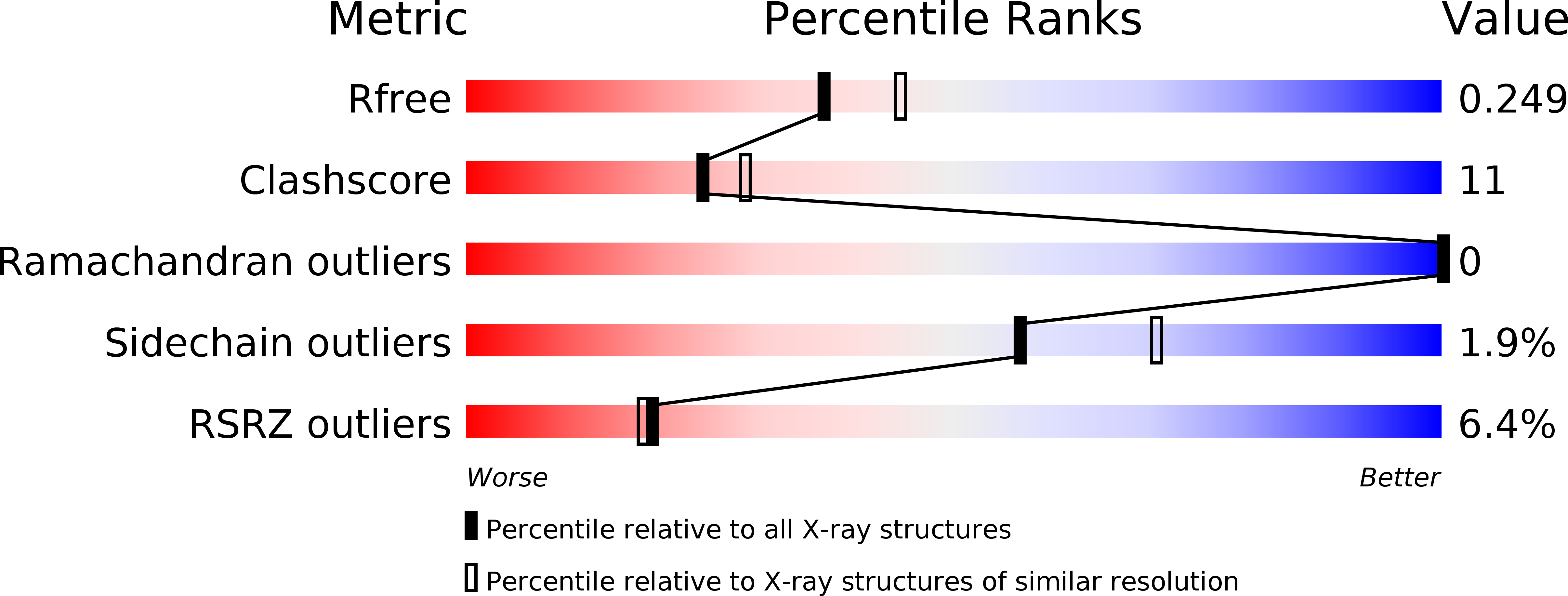
1UAM image
Deposition Date 2003-03-11
Release Date 2003-06-17
Last Version Date 2023-12-27
Entry Detail
PDB ID:
1UAM
Keywords:
Title:
Crystal structure of tRNA(m1G37)methyltransferase: Insight into tRNA recognition
Biological Source:
Source Organism:
Host Organism:
Method Details:
Experimental Method:
Resolution:
2.20 Å
R-Value Free:
0.26
R-Value Work:
0.22
R-Value Observed:
0.22
Space Group:
H 3 2
Macromolecular Entities
Polymer Type:polypeptide(L)
Molecule:tRNA (Guanine-N(1)-)-methyltransferase
Gene (Uniprot):trmD
Chain IDs:A
Chain Length:274
Number of Molecules:1
Biological Source:Haemophilus influenzae
Primary Citation
Crystal structure of tRNA(m(1)G37)methyltransferase: insights into tRNA recognition
EMBO J. 22 2593 2603 (2003)
PMID: 12773376 DOI: 10.1093/emboj/cdg269

Abstact

tRNA(m(1)G37)methyltransferase (TrmD) catalyzes the transfer of a methyl group from S-adenosyl-L- methionine (AdoMet) to G(37) within a subset of bacterial tRNA species, which have a G residue at the 36th position. The modified guanosine is adjacent to and 3' of the anticodon and is essential for the maintenance of the correct reading frame during translation. Here we report four crystal structures of TrmD from Haemophilus influenzae, as binary complexes with either AdoMet or S-adenosyl-L-homocysteine (AdoHcy), as a ternary complex with AdoHcy and phosphate, and as an apo form. This first structure of TrmD indicates that it functions as a dimer. It also suggests the binding mode of G(36)G(37) in the active site of TrmD and the catalytic mechanism. The N-terminal domain has a trefoil knot, in which AdoMet or AdoHcy is bound in a novel, bent conformation. The C-terminal domain shows structural similarity to trp repressor. We propose a plausible model for the TrmD(2)-tRNA(2) complex, which provides insights into recognition of the general tRNA structure by TrmD.

Legend

Protein

Chemical

Disease

Primary Citation of related structures