1UA7 image
Deposition Date 2003-03-03
Release Date 2004-05-18
Last Version Date 2023-12-27
Entry Detail
PDB ID:
1UA7
Keywords:
Title:
Crystal Structure Analysis of Alpha-Amylase from Bacillus Subtilis complexed with Acarbose
Biological Source:
Source Organism:
Host Organism:
Method Details:
Experimental Method:
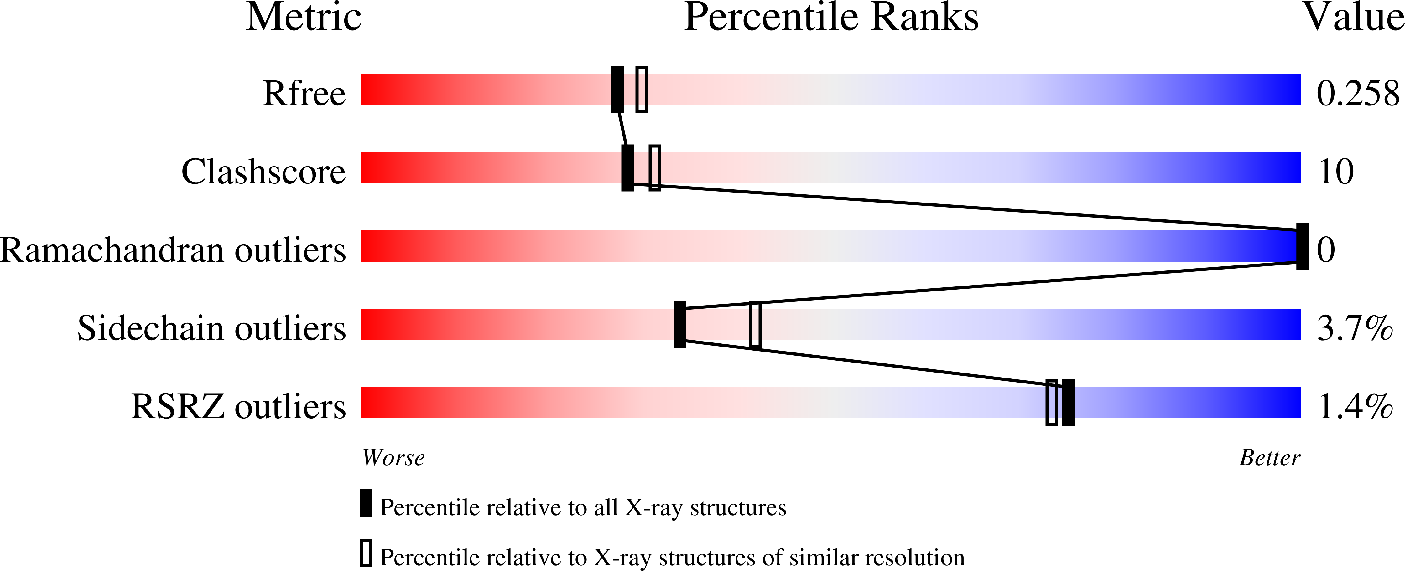
Resolution:
2.21 Å
R-Value Free:
0.26
R-Value Work:
0.20
R-Value Observed:
0.20
Space Group:
P 21 21 21
Macromolecular Entities
Structures with similar UniProt ID
Protein Blast
Polymer Type:polypeptide(L)
Molecule:Alpha-amylase
Gene (Uniprot):amyE
Mutagens:N356Q
Chain IDs:A
Chain Length:422
Number of Molecules:1
Biological Source:Bacillus subtilis
Primary Citation

Abstact

The crystal structure of Bacillus subtilis alpha-amylase, in complex with the pseudotetrasaccharide inhibitor acarbose, revealed an hexasaccharide in the active site as a result of transglycosylation. After comparison with the known structure of the catalytic-site mutant complexed with the native substrate maltopentaose, it is suggested that the present structure represents a mimic intermediate in the initial stage of the catalytic process.

Legend

Protein

Chemical

Disease

Primary Citation of related structures
Feedback Form
Name
Email
Institute
Feedback