1UA2 image
Deposition Date 2004-08-11
Release Date 2004-12-07
Last Version Date 2024-11-20
Entry Detail
PDB ID:
1UA2
Title:
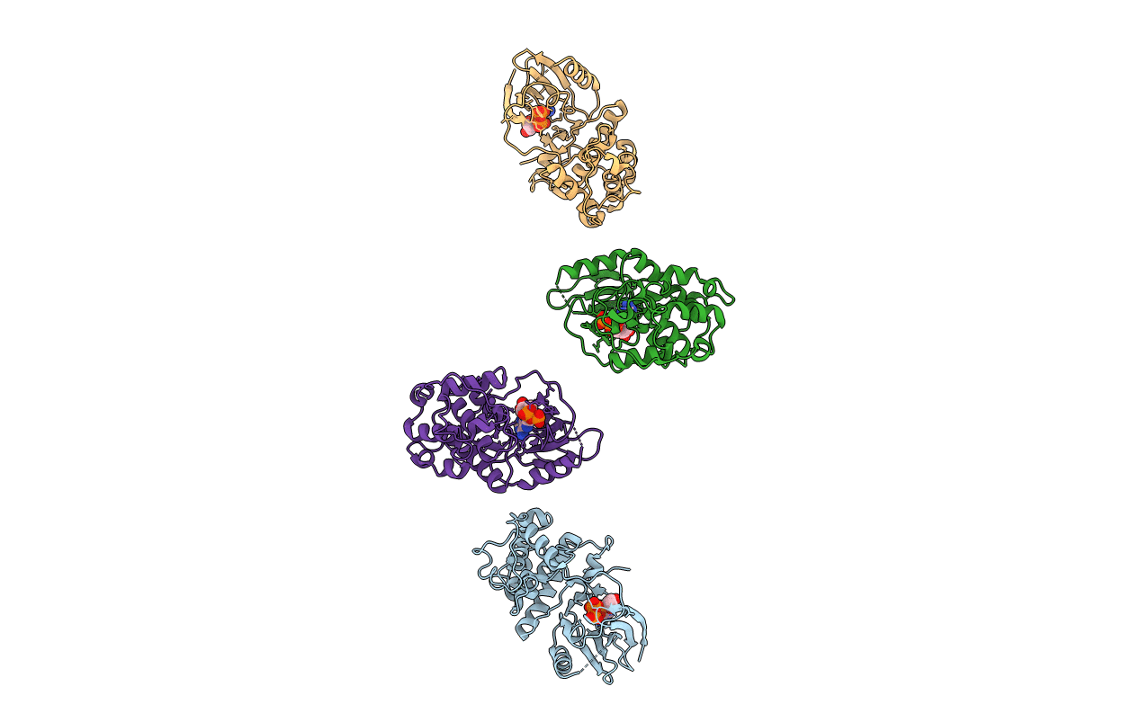
Crystal Structure of Human CDK7
Biological Source:
Source Organism:
Homo sapiens (Taxon ID: 9606)
Host Organism:
Method Details:
Experimental Method:
Resolution:
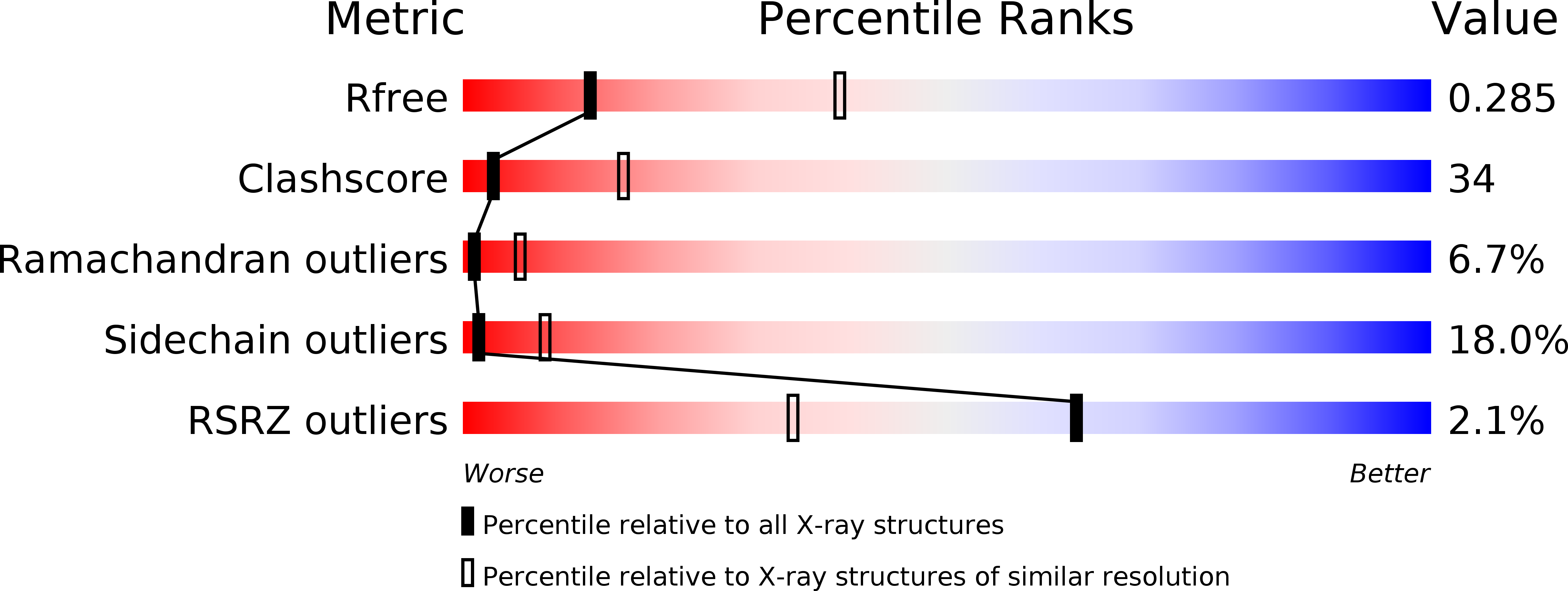
3.02 Å
R-Value Free:
0.28
R-Value Work:
0.21
R-Value Observed:
0.21
Space Group:
P 1 21 1
Macromolecular Entities
Polymer Type:polypeptide(L)
Molecule:Cell division protein kinase 7
Gene (Uniprot):CDK7
Chain IDs:A, B, C, D
Chain Length:346
Number of Molecules:4
Biological Source:Homo sapiens
Modified Residue
Compound ID Chain ID Parent Comp ID Details 2D Image
TPO A THR PHOSPHOTHREONINE
Ligand Molecules
Primary Citation
The Crystal Structure of Human CDK7 and Its Protein Recognition Properties
Structure 12 2067 2079 (2004)
PMID: 15530371 DOI: 10.1016/j.str.2004.08.013

Abstact

CDK7, a member of the cyclin-dependent protein kinase family, regulates the activities of other CDKs through phosphorylation on their activation segment and hence contributes to control of the eukaryotic cell cycle. CDK7 also assists in the regulation of transcription as part of the transcription factor TFIIH complex. For maximum activity and stability, CDK7 requires phosphorylation, association with cyclin H, and association with a third protein, MAT1. We have determined the crystal structure of human CDK7 in complex with ATP at 3 A resolution. The kinase is in the inactive conformation, similar to that observed for inactive CDK2. The activation segment is phosphorylated at Thr170 and is in a defined conformation that differs from that in phospho-CDK2 and phospho-CDK2/cyclin A. The functional properties of the enzyme against CDK2 and CTD as substrates are characterized through kinase assays. Experiments confirm that CDK7 is not a substrate for kinase-associated phosphatase.

Legend

Protein

Chemical

Disease

Primary Citation of related structures