1U9T image
Deposition Date 2004-08-10
Release Date 2005-10-25
Last Version Date 2024-02-14
Entry Detail
PDB ID:
1U9T
Keywords:
Title:
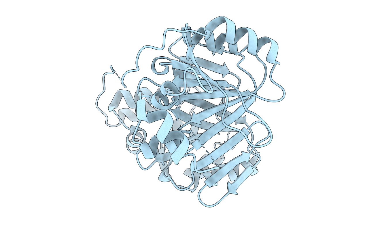
Crystal Structure Analysis of ChuS, an E. coli Heme Oxygenase
Biological Source:
Source Organism:
Method Details:
Experimental Method:
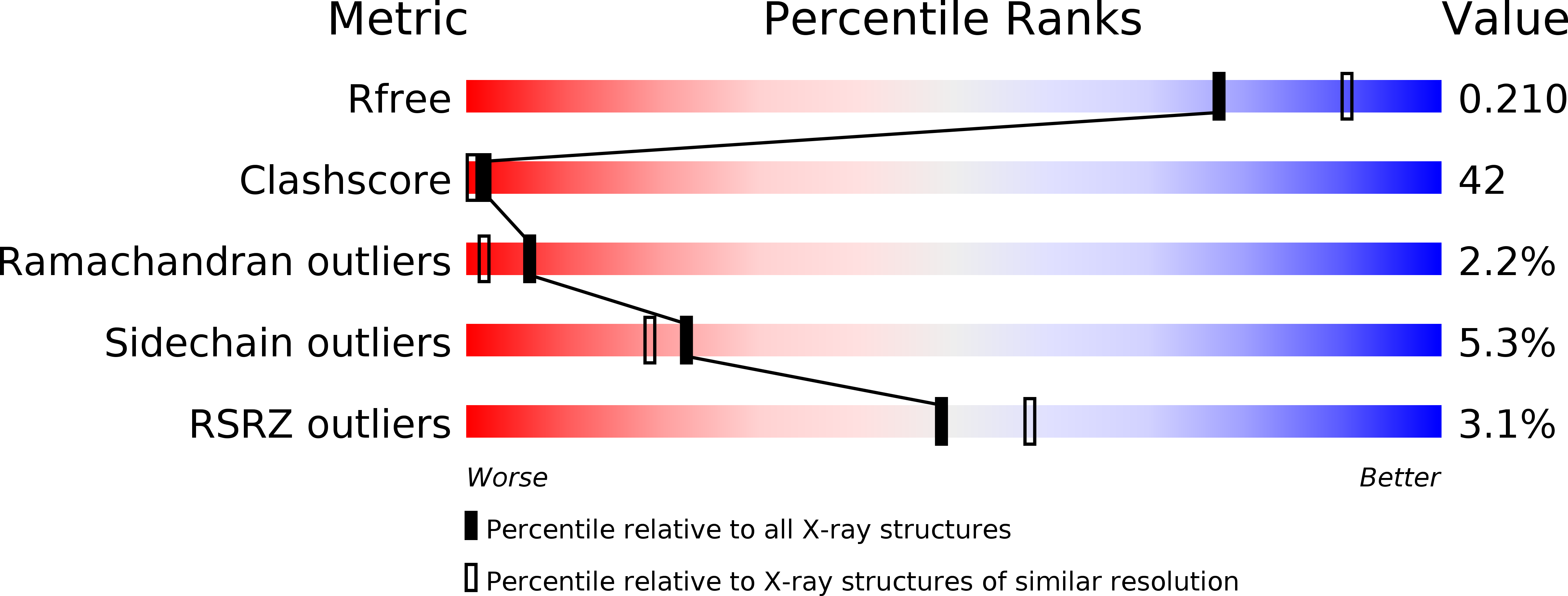
Resolution:
2.16 Å
R-Value Free:
0.28
R-Value Work:
0.20
R-Value Observed:
0.20
Space Group:
P 1 21 1
Macromolecular Entities
Polymer Type:polypeptide(L)
Molecule:putative heme/hemoglobin transport protein
Gene (Uniprot):ECs_4379
Chain IDs:A
Chain Length:354
Number of Molecules:1
Biological Source:Escherichia coli
Primary Citation
Identification of an Escherichia coli O157:H7 heme oxygenase with tandem functional repeats
Proc.Natl.Acad.Sci.Usa 102 16955 16960 (2005)
PMID: 16275907 DOI: 10.1073/pnas.0504289102

Abstact

Heme oxygenases (HOs) catalyze the oxidation of heme to biliverdin, carbon monoxide (CO), and free iron. Iron acquisition is critical for invading microorganisms to enable survival and growth. Here we report the crystal structure of ChuS, which displays a previously uncharacterized fold and is unique compared with other characterized HOs. Despite only 19% sequence identity between the N- and C-terminal halves, these segments of ChuS represent a structural duplication, with a root-mean-square deviation of 2.1 A between the two repeats. ChuS is capable of using ascorbic acid or cytochrome P450 reductase-NADPH as electron sources for heme oxygenation. CO detection confirmed that ChuS is a HO, and we have identified it in pathogenic Escherichia coli O157:H7. Based on sequence analysis, this HO is present in many bacteria, although not in the E. coli K-12 strain. The N- and C-terminal halves of ChuS are each a functional HO.

Legend

Protein

Chemical

Disease

Primary Citation of related structures