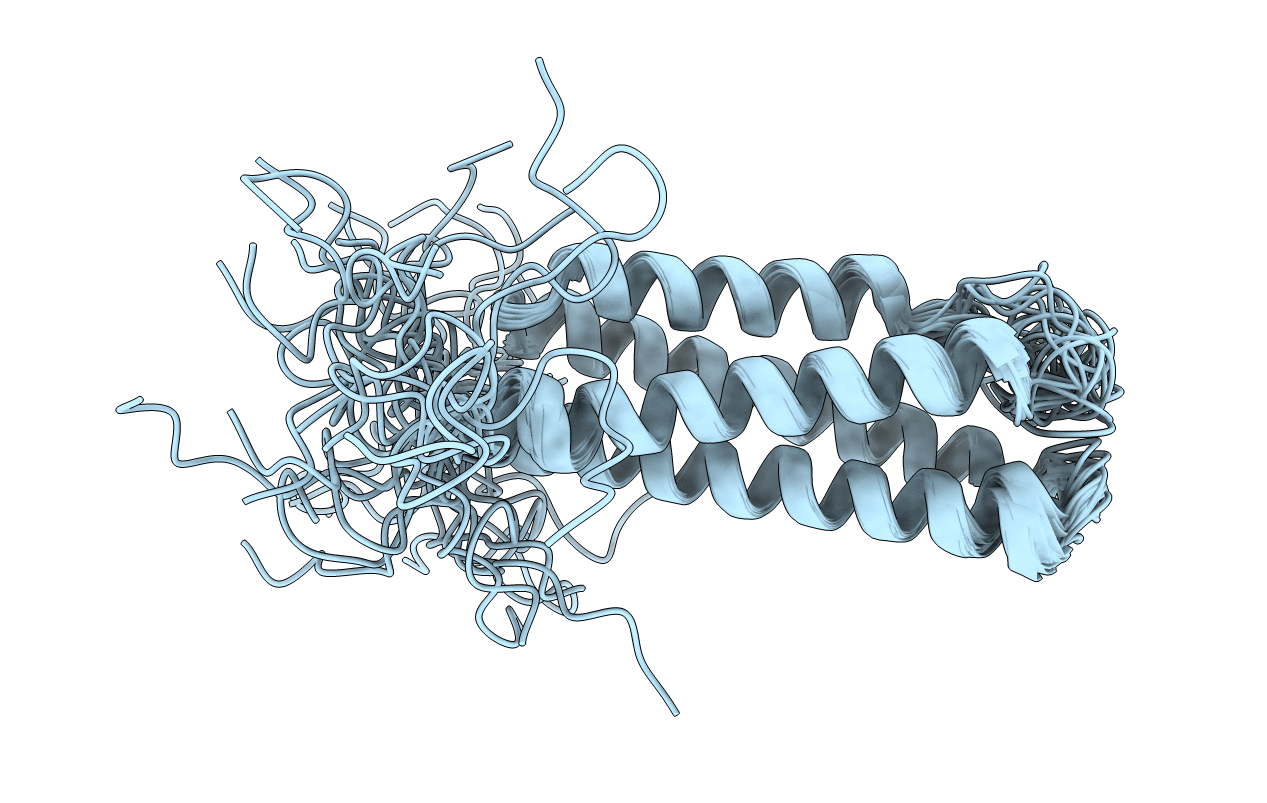
1U89 image
Deposition Date 2004-08-05
Release Date 2005-01-18
Last Version Date 2024-05-29
Entry Detail
PDB ID:
1U89
Title:
Solution structure of VBS2 fragment of talin
Biological Source:
Source Organism:
Mus musculus (Taxon ID: 10090)
Method Details:
Experimental Method:
Conformers Calculated:
100
Conformers Submitted:
20
Selection Criteria:
structures with the lowest energy
Macromolecular Entities
Structures with similar UniProt ID
Protein Blast
Polymer Type:polypeptide(L)
Molecule:Talin 1
Gene (Uniprot):Tln1
Chain IDs:A
Chain Length:139
Number of Molecules:1
Biological Source:Mus musculus
Ligand Molecules
Primary Citation
A vinculin binding domain from the talin rod unfolds to form a complex with the vinculin head.
Structure 13 65 74 (2005)
PMID: 15642262 DOI: 10.1016/j.str.2004.11.006

Abstact

The cytoskeletal protein talin plays a key role in activating integrins and in coupling them to the actin cytoskeleton. Its N-terminal globular head, which binds beta integrins, is linked to an extended rod having a C-terminal actin binding site and several vinculin binding sites (VBSs). The NMR structure of residues 755-889 of the rod (containing a VBS) is shown to be an amphipathic four-helix bundle with a left-handed topology. A talin peptide corresponding to the VBS binds the vinculin head; the X-ray crystallographic structure of this complex shows that the residues which interact with vinculin are buried in the hydrophobic core of the talin fragment. NMR shows that the interaction involves a major structural change in the talin fragment, including unfolding of one of its helices, making the VBS accessible to vinculin. Interestingly, the talin 755-889 fragment binds more than one vinculin head molecule, suggesting that the talin rod may contain additional as yet unrecognized VBSs.

Legend

Protein

Chemical

Disease

Primary Citation of related structures