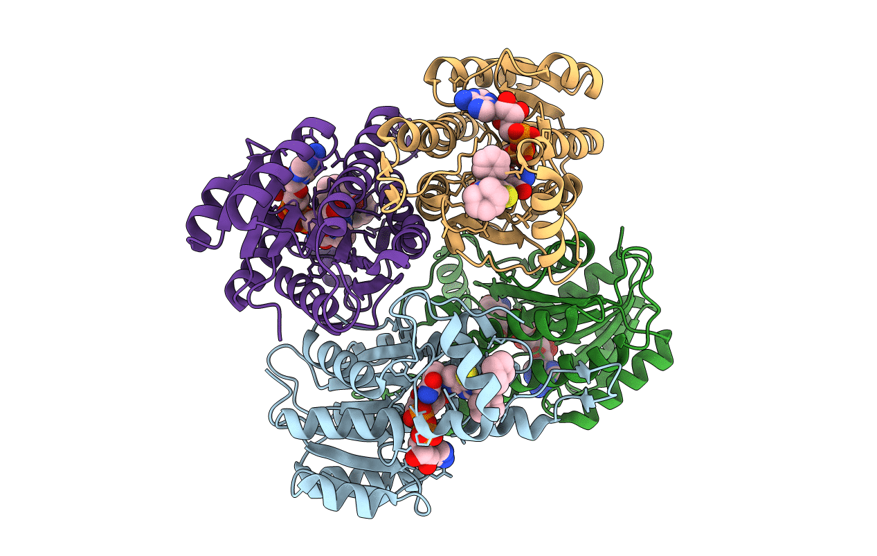
1U7T image
Deposition Date 2004-08-04
Release Date 2004-10-05
Last Version Date 2024-04-03
Entry Detail
PDB ID:
1U7T
Keywords:
Title:
Crystal Structure of ABAD/HSD10 with a Bound Inhibitor
Biological Source:
Source Organism:
Homo sapiens (Taxon ID: 9606)
Host Organism:
Method Details:
Experimental Method:
Resolution:
2.00 Å
R-Value Free:
0.26
R-Value Work:
0.21
R-Value Observed:
0.21
Space Group:
C 1 2 1
Macromolecular Entities
Polymer Type:polypeptide(L)
Molecule:3-hydroxyacyl-CoA dehydrogenase type II
Gene (Uniprot):HSD17B10
Mutations:C214R
Chain IDs:A, B, C, D
Chain Length:261
Number of Molecules:4
Biological Source:Homo sapiens
Primary Citation
Crystal structure of human ABAD/HSD10 with a bound inhibitor: implications for design of Alzheimer's disease therapeutics
J.Mol.Biol. 342 943 952 (2004)
PMID: 15342248 DOI: 10.1016/j.jmb.2004.07.071

Abstact

The enzyme 17beta-hydroxysteroid dehydrogenase type 10 (HSD10), also known as amyloid beta-peptide-binding alcohol dehydrogenase (ABAD), has been implicated in the development of Alzheimer's disease. This protein, a member of the short-chain dehydrogenase/reductase family of enzymes, has been shown to bind beta-amyloid and to participate in beta-amyloid neurotoxicity. We have determined the crystal structure of human ABAD/HSD10 complexed with NAD(+) and an inhibitory small molecule. The inhibitor occupies the substrate-binding site and forms a covalent adduct with the NAD(+) cofactor. The crystal structure provides a basis for the design of potent, highly specific ABAD/HSD10 inhibitors with potential application in the treatment of Alzheimer's disease.

Legend

Protein

Chemical

Disease

Primary Citation of related structures