1U7L image
Deposition Date 2004-08-04
Release Date 2004-11-23
Last Version Date 2024-03-20
Entry Detail
PDB ID:
1U7L
Title:
Crystal Structure of subunit C (vma5p) of the yeast V-ATPase
Biological Source:
Source Organism:
Host Organism:
Method Details:
Experimental Method:
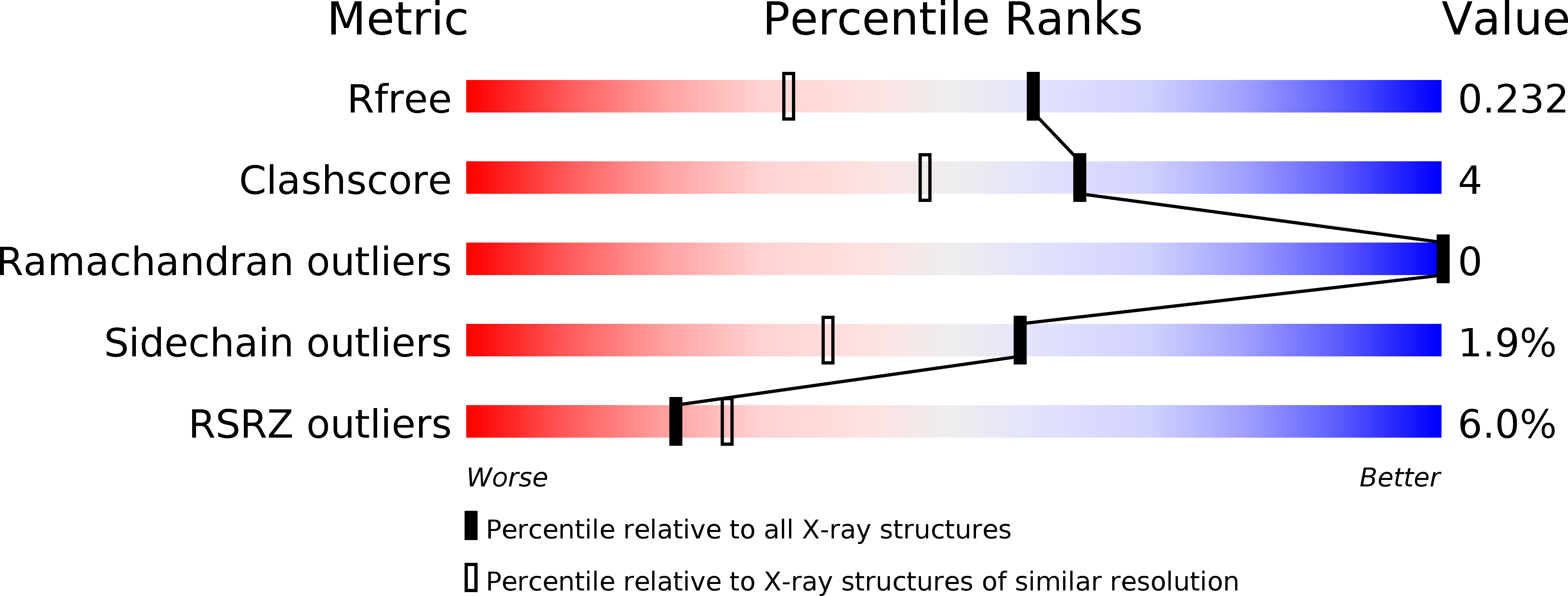
Resolution:
1.75 Å
R-Value Free:
0.23
R-Value Work:
0.20
R-Value Observed:
0.21
Space Group:
P 43 21 2
Macromolecular Entities
Structures with similar UniProt ID
Protein Blast
Polymer Type:polypeptide(L)
Molecule:Vacuolar ATP synthase subunit C
Gene (Uniprot):VMA5
Chain IDs:A
Chain Length:392
Number of Molecules:1
Biological Source:Saccharomyces cerevisiae
Ligand Molecules
Primary Citation
Crystal structure of yeast V-ATPase subunit C reveals its stator function
Embo Rep. 5 1148 1152 (2004)
PMID: 15540116 DOI: 10.1038/sj.embor.7400294

Abstact

Vacuolar H(+)-ATPase (V-ATPase) has a crucial role in the vacuolar system of eukaryotic cells. It provides most of the energy required for transport systems that utilize the proton-motive force that is generated by ATP hydrolysis. Some, but not all, of the V-ATPase subunits are homologous to those of F-ATPase and the nonhomologous subunits determine the unique features of V-ATPase. We determined the crystal structure of V-ATPase subunit C (Vma5p), which does not show any homology with F-ATPase subunits, at 1.75 A resolution. The structural features suggest that subunit C functions as a flexible stator that holds together the catalytic and membrane sectors of the enzyme. A second crystal form that was solved at 2.9 A resolution supports the flexible nature of subunit C. These structures provide a framework for exploring the unique mechanistic features of V-ATPases.

Legend

Protein

Chemical

Disease

Primary Citation of related structures
Feedback Form
Name
Email
Institute
Feedback