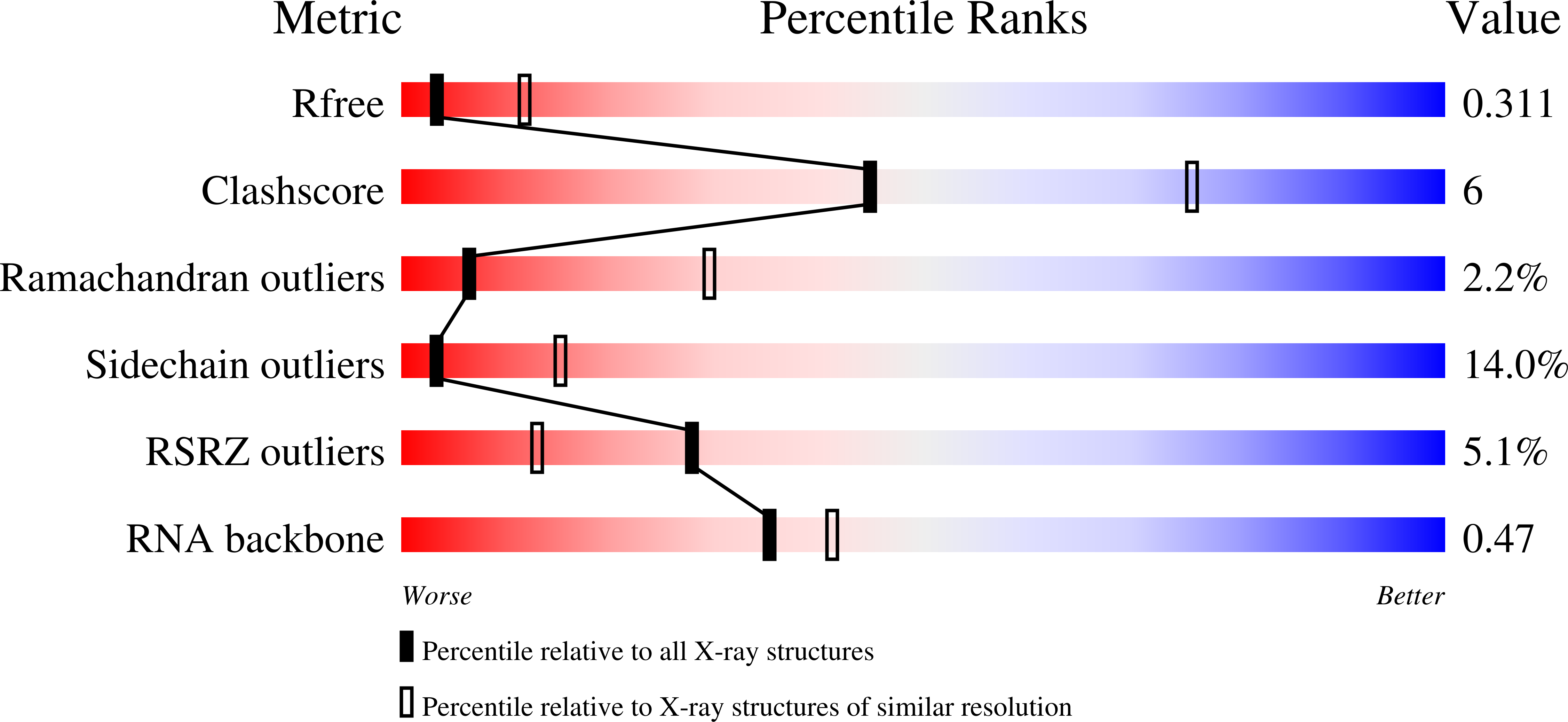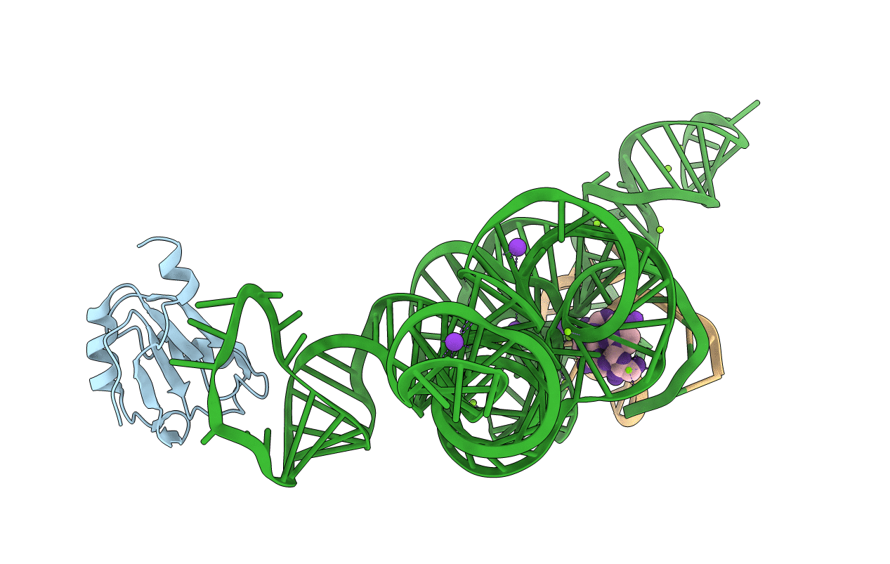
1U6B image
Deposition Date 2004-07-29
Release Date 2004-08-10
Last Version Date 2024-02-14
Entry Detail
PDB ID:
1U6B
Title:
CRYSTAL STRUCTURE OF A SELF-SPLICING GROUP I INTRON WITH BOTH EXONS
Biological Source:
Source Organism:
Homo sapiens (Taxon ID: 9606)
Host Organism:
Method Details:
Experimental Method:
Resolution:
3.10 Å
R-Value Free:
0.27
R-Value Work:
0.24
R-Value Observed:
0.24
Space Group:
P 41 2 2
Macromolecular Entities
Structures with similar UniProt ID
Protein Blast
Polymer Type:polypeptide(L)
Molecule:U1 small nuclear ribonucleoprotein A
Gene (Uniprot):SNRPA
Mutagens:Y31H,Q36R
Chain IDs:D (auth: A)
Chain Length:98
Number of Molecules:1
Biological Source:Homo sapiens
Polymer Type:polyribonucleotide
Molecule:197-MER
Chain IDs:A (auth: B)
Chain Length:197
Number of Molecules:1
Biological Source:
Modified Residue
Compound ID Chain ID Parent Comp ID Details 2D Image
5MU C U 5-METHYLURIDINE 5'-MONOPHOSPHATE
A23 A A ?
GTP A G GUANOSINE-5'-TRIPHOSPHATE
Primary Citation

Abstact

The discovery of the RNA self-splicing group I intron provided the first demonstration that not all enzymes are proteins. Here we report the X-ray crystal structure (3.1-A resolution) of a complete group I bacterial intron in complex with both the 5'- and the 3'-exons. This complex corresponds to the splicing intermediate before the exon ligation step. It reveals how the intron uses structurally unprecedented RNA motifs to select the 5'- and 3'-splice sites. The 5'-exon's 3'-OH is positioned for inline nucleophilic attack on the conformationally constrained scissile phosphate at the intron-3'-exon junction. Six phosphates from three disparate RNA strands converge to coordinate two metal ions that are asymmetrically positioned on opposing sides of the reactive phosphate. This structure represents the first splicing complex to include a complete intron, both exons and an organized active site occupied with metal ions.

Legend

Protein

Chemical

Disease

Primary Citation of related structures