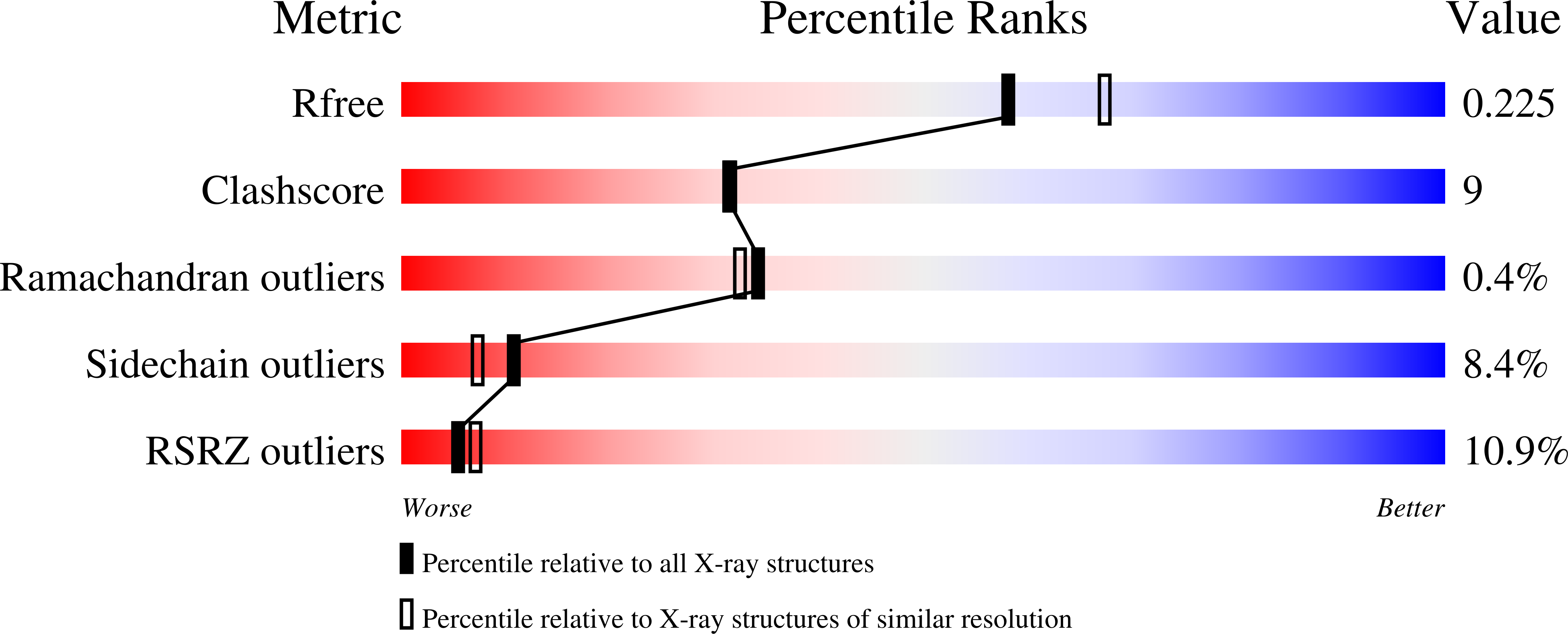
1U5G image
Deposition Date 2004-07-27
Release Date 2005-07-26
Last Version Date 2023-08-23
Entry Detail
PDB ID:
1U5G
Title:
Crystal Structure of the PH Domain of SKAP-Hom
Biological Source:
Source Organism:
Mus musculus (Taxon ID: 10090)
Method Details:
Experimental Method:
Resolution:
2.10 Å
R-Value Free:
0.27
R-Value Work:
0.21
R-Value Observed:
0.21
Space Group:
P 21 21 21
Macromolecular Entities
Polymer Type:polypeptide(L)
Molecule:Src-associated adaptor protein
Mutations:S101G, D102S
Chain IDs:A, B, C, D
Chain Length:122
Number of Molecules:4
Biological Source:Mus musculus
Primary Citation
The Skap-hom dimerization and PH domains comprise a 3'-phosphoinositide-gated molecular switch.
Mol.Cell 32 564 575 (2008)
PMID: 19026786 DOI: 10.1016/j.molcel.2008.09.022

Abstact

PH domains, by binding to phosphoinositides, often serve as membrane-targeting modules. Using crystallographic, biochemical, and cell biological approaches, we have uncovered a mechanism that the integrin-signaling adaptor Skap-hom uses to mediate cytoskeletal interactions. Skap-hom is a homodimer containing an N-terminal four-helix bundle dimerization domain, against which its two PH domains pack in a conformation incompatible with phosphoinositide binding. The isolated PH domains bind PI[3,4,5]P(3), and mutations targeting the dimerization domain or the PH domain's PI[3,4,5]P(3)-binding pocket prevent Skap-hom localization to ruffles. Targeting is retained when the PH domain is deleted or by combined mutation of the PI[3,4,5]P(3)-binding pocket and the PH/dimerization domain interface. Thus, the dimerization and PH domain form a PI[3,4,5]P(3)-responsive molecular switch that controls Skap-hom function.

Legend

Protein

Chemical

Disease

Primary Citation of related structures