1U55 image
Deposition Date 2004-07-27
Release Date 2004-08-31
Last Version Date 2023-08-23
Entry Detail
PDB ID:
1U55
Title:
Crystal structure of an oxygen binding H-NOX domain related to soluble guanylate cyclases (oxygen complex)
Biological Source:
Source Organism:
Host Organism:
Method Details:
Experimental Method:
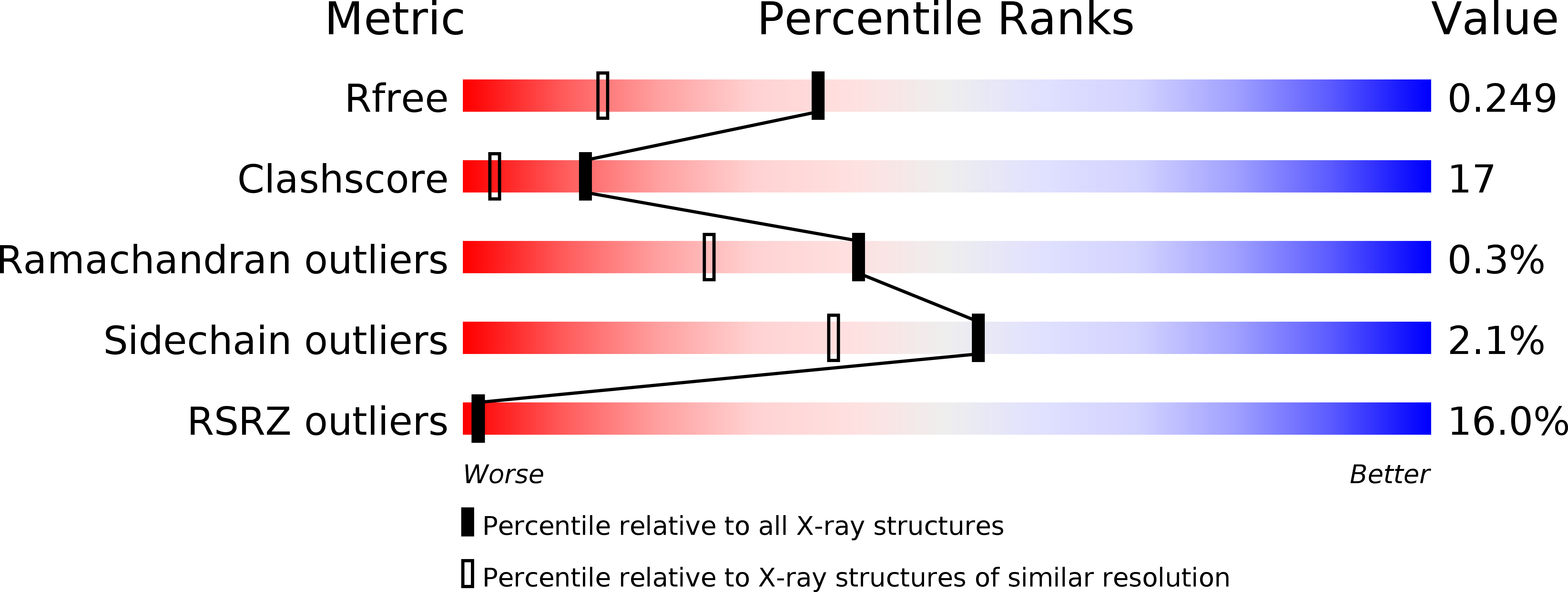
Resolution:
1.77 Å
R-Value Free:
0.25
R-Value Work:
0.22
Space Group:
C 1 2 1
Macromolecular Entities
Polymer Type:polypeptide(L)
Molecule:Heme-based Methyl-accepting Chemotaxis Protein
Gene (Uniprot):Tar4
Chain IDs:A, B
Chain Length:188
Number of Molecules:2
Biological Source:Thermoanaerobacter tengcongensis
Primary Citation
Crystal structure of an oxygen-binding heme domain related to soluble guanylate cyclases.
Proc.Natl.Acad.Sci.Usa 101 12854 12859 (2004)
PMID: 15326296 DOI: 10.1073/pnas.0405188101

Abstact

Soluble guanylate cyclases are nitric oxide-responsive signaling proteins in which the nitric oxide sensor is a heme-binding domain of unknown structure that we have termed the heme-NO and oxygen binding (H-NOX) domain. H-NOX domains are also found in bacteria, either as isolated domains, or are fused through a membrane-spanning region to methyl-accepting chemotaxis proteins. We have determined the crystal structure of an oxygen-binding H-NOX domain of one such signaling protein from the obligate anaerobe Thermoanaerobacter tengcongensis at 1.77-angstroms resolution, revealing a protein fold unrelated to known structures. Particularly striking is the structure of the protoporphyrin IX group, which is distorted from planarity to an extent not seen before in protein-bound heme groups. Comparison of the structure of the H-NOX domain in two different crystal forms suggests a mechanism whereby alteration in the degree of distortion of the heme group is coupled to changes on the molecular surface of the H-NOX domain and potentially to changes in intermolecular interactions.

Legend

Protein

Chemical

Disease

Primary Citation of related structures