Deposition Date
2004-07-21
Release Date
2004-08-31
Last Version Date
2024-04-03
Entry Detail
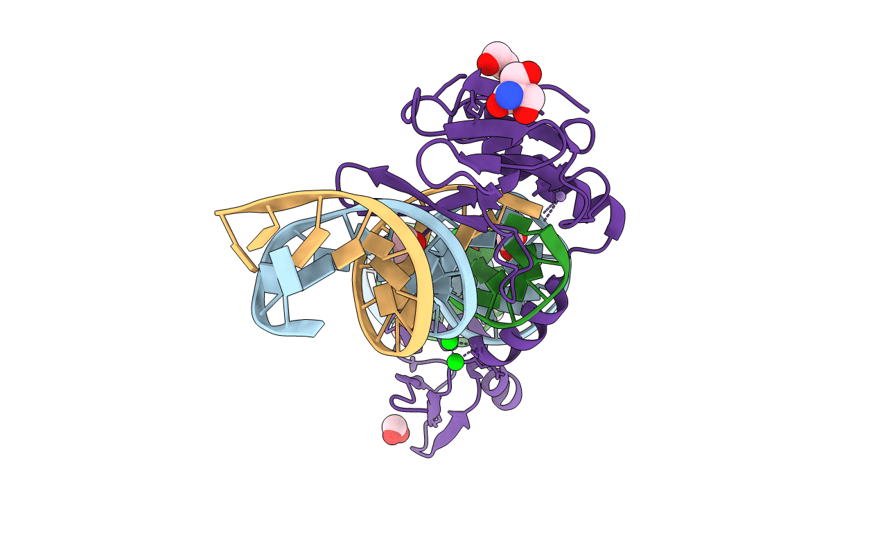
PDB ID:
1U3E
Keywords:
Title:
DNA binding and cleavage by the HNH homing endonuclease I-HmuI
Biological Source:
Source Organism(s):
Bacillus phage SPO1 (Taxon ID: 10685)
Expression System(s):
Method Details:
Experimental Method:
Resolution:
2.92 Å
R-Value Free:
0.24
R-Value Work:
0.21
R-Value Observed:
0.22
Space Group:
I 2 2 2