1U3C image
Entry Detail
PDB ID:
1U3C
Title:
Crystal Structure of the PHR domain of Cryptochrome 1 from Arabidopsis thaliana
Biological Source:
Source Organism:
Host Organism:
PDB Version:
Deposition Date:
2004-07-21
Release Date:
2004-08-24
Method Details:
Experimental Method:
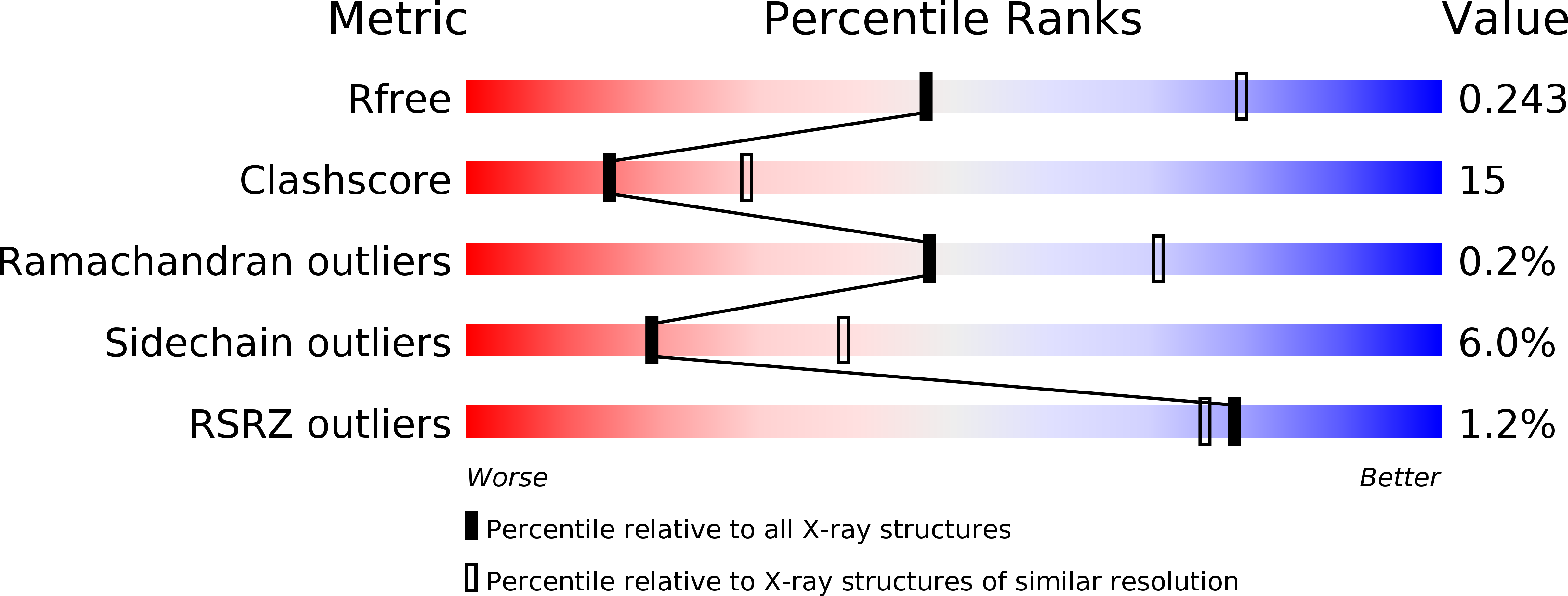
Resolution:
2.60 Å
R-Value Free:
0.25
R-Value Work:
0.20
R-Value Observed:
0.20
Space Group:
P 63 2 2
Macromolecular Entities
Polymer Type:polypeptide(L)
Description:Cryptochrome 1 apoprotein
Chain IDs:A
Chain Length:509
Number of Molecules:1
Biological Source:Arabidopsis thaliana
Primary Citation
Structure of the photolyase-like domain of cryptochrome 1 from Arabidopsis thaliana.
Proc.Natl.Acad.Sci.USA 101 12142 12147 (2004)
PMID: 15299148 DOI: 10.1073/pnas.0404851101

Abstact

Signals generated by cryptochrome (CRY) blue-light photoreceptors are responsible for a variety of developmental and circadian responses in plants. The CRYs are also identified as circadian blue-light photoreceptors in Drosophila and components of the mammalian circadian clock. These flavoproteins all have an N-terminal domain that is similar to photolyase, and most have an additional C-terminal domain of variable length. We present here the crystal structure of the photolyase-like domain of CRY-1 from Arabidopsis thaliana. The structure reveals a fold that is very similar to photolyase, with a single molecule of FAD noncovalently bound to the protein. The surface features of the protein and the dissimilarity of a surface cavity to that of photolyase account for its lack of DNA-repair activity. Previous in vitro experiments established that the photolyase-like domain of CRY-1 can bind Mg.ATP, and we observe a single molecule of an ATP analog bound in the aforementioned surface cavity, near the bound FAD cofactor. The structure has implications for the signaling mechanism of CRY blue-light photoreceptors.

Legend

Protein

Chemical

Disease

Primary Citation of related structures