1U2X image
Deposition Date 2004-07-20
Release Date 2004-09-14
Last Version Date 2024-10-30
Entry Detail
PDB ID:
1U2X
Keywords:
Title:
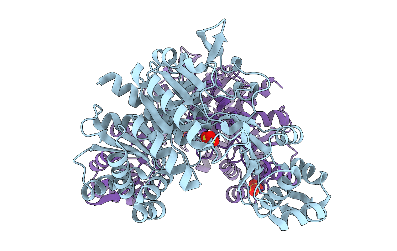
Crystal Structure of a Hypothetical ADP-dependent Phosphofructokinase from Pyrococcus horikoshii OT3
Biological Source:
Source Organism:
Method Details:
Experimental Method:
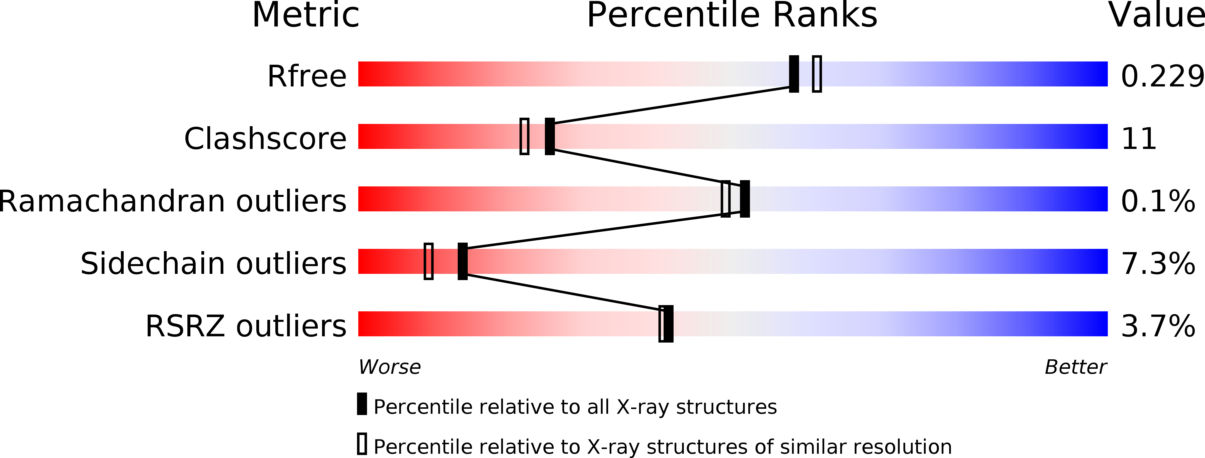
Resolution:
2.00 Å
R-Value Free:
0.22
R-Value Work:
0.17
R-Value Observed:
0.17
Space Group:
P 1 21 1
Macromolecular Entities
Polymer Type:polypeptide(L)
Molecule:ADP-specific phosphofructokinase
Gene (Uniprot):pfkC
Chain IDs:A, B
Chain Length:474
Number of Molecules:2
Biological Source:Pyrococcus horikoshii
Modified Residue
Compound ID Chain ID Parent Comp ID Details 2D Image
MSE A MET SELENOMETHIONINE
Ligand Molecules
Primary Citation
ADP-dependent 6-phosphofructokinase from Pyrococcus horikoshii OT3: structure determination and biochemical characterization of PH1645.
J.Biol.Chem. 284 22664 22671 (2009)
PMID: 19553681 DOI: 10.1074/jbc.M109.012401

Abstact

Some hyperthermophilic archaea use a modified glycolytic pathway that employs an ADP-dependent glucokinase (ADP-GK) and an ADP-dependent phosphofructokinase (ADP-PFK) or, in the case of Methanococcus jannaschii, a bifunctional ADP-dependent glucophosphofructokinase (ADP-GK/PFK). The crystal structures of three ADP-GKs have been determined. However, there is no structural information available for ADP-PFKs or the ADP-GK/PFK. Here, we present the first crystal structure of an ADP-PFK from Pyrococcus horikoshii OT3 (PhPFK) in both apo- and AMP-bound forms determined to 2.0-A and 1.9-A resolution, respectively, along with biochemical characterization of the enzyme. The overall structure of PhPFK maintains a similar large and small alpha/beta domain structure seen in the ADP-GK structures. A large conformational change accompanies binding of phosphoryl donor, acceptor, or both, in all members of the ribokinase superfamily characterized thus far, which is believed to be critical to enzyme function. Surprisingly, no such conformational change was observed in the AMP-bound PhPFK structure compared with the apo structure. Through comprehensive site-directed mutagenesis of the substrate binding pocket we identified residues that were critical for both substrate recognition and the phosphotransfer reaction. The catalytic residues and many of the substrate binding residues are conserved between PhPFK and ADP-GKs; however, four key residues differ in the sugar-binding pocket, which we have shown determine the sugar-binding specificity. Using these results we were able to engineer a mutant PhPFK that mimics the ADP-GK/PFK and is able to phosphorylate both fructose 6-phosphate and glucose.

Legend

Protein

Chemical

Disease

Primary Citation of related structures