Deposition Date
2004-07-19
Release Date
2005-07-05
Last Version Date
2023-10-25
Entry Detail
PDB ID:
1U2H
Keywords:
Title:
X-ray Structure of the N-terminally truncated human APEP-1
Biological Source:
Source Organism(s):
Homo sapiens (Taxon ID: 9606)
Expression System(s):
Method Details:
Experimental Method:
Resolution:
0.96 Å
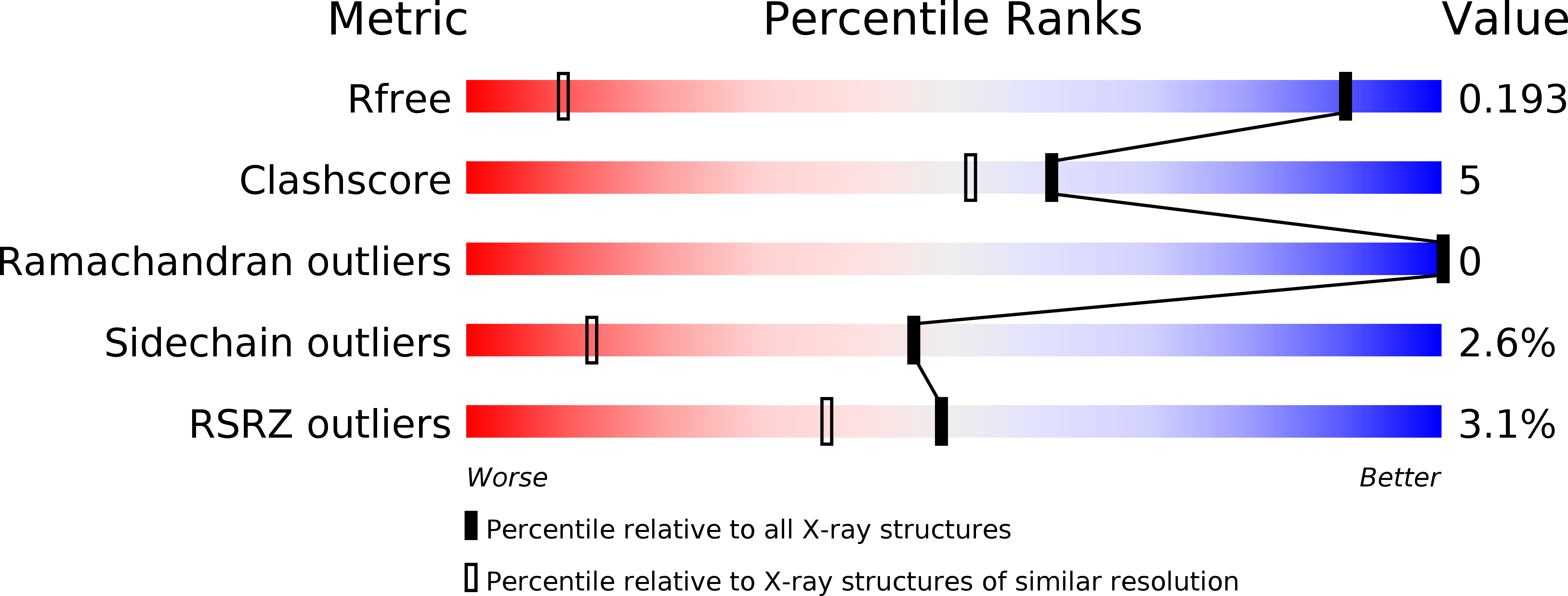
R-Value Free:
0.17
R-Value Work:
0.16
R-Value Observed:
0.16
Space Group:
C 1 2 1