Deposition Date
2004-07-16
Release Date
2005-01-18
Last Version Date
2023-08-23
Entry Detail
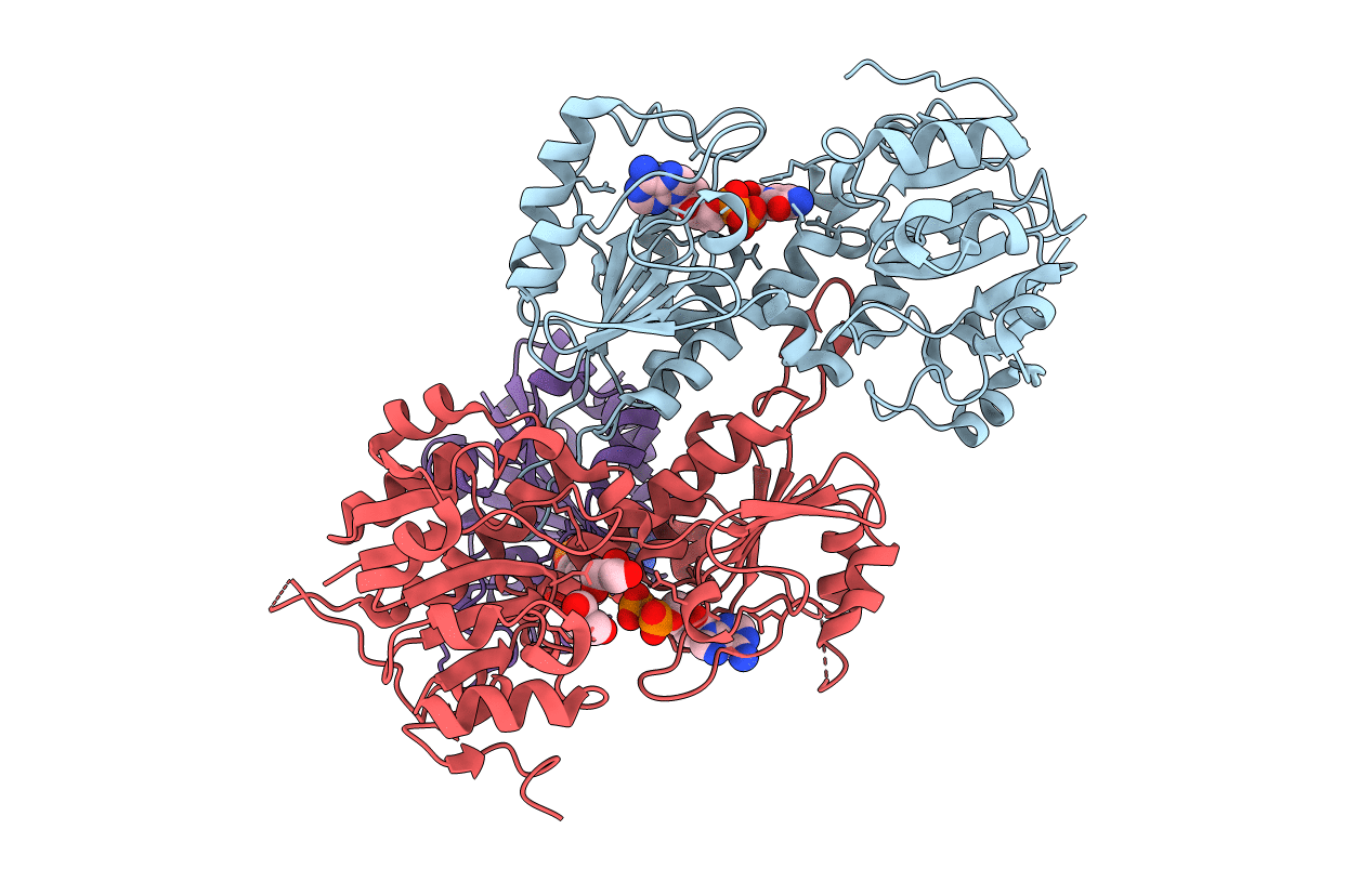
PDB ID:
1U28
Keywords:
Title:
R. rubrum transhydrogenase asymmetric complex (dI.NAD+)2(dIII.NADP+)1
Biological Source:
Source Organism(s):
Rhodospirillum rubrum (Taxon ID: 1085)
Expression System(s):
Method Details:
Experimental Method:
Resolution:
2.30 Å
R-Value Free:
0.27
R-Value Work:
0.24
Space Group:
P 21 21 21