Deposition Date
2004-07-15
Release Date
2004-10-12
Last Version Date
2024-10-09
Entry Detail
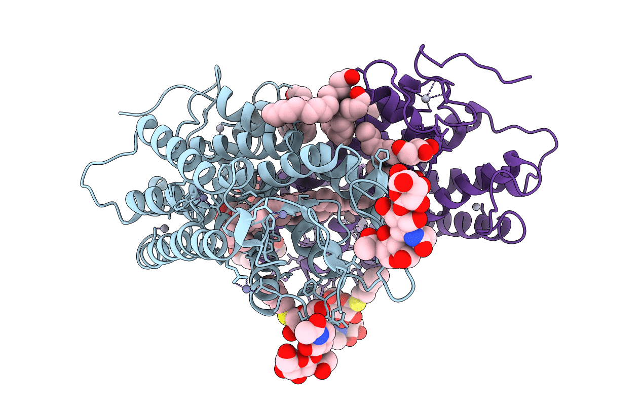
PDB ID:
1U19
Keywords:
Title:
Crystal Structure of Bovine Rhodopsin at 2.2 Angstroms Resolution
Biological Source:
Source Organism(s):
Bos taurus (Taxon ID: 9913)
Method Details:
Experimental Method:
Resolution:
2.20 Å
R-Value Free:
0.22
R-Value Work:
0.2
Space Group:
P 41