1U00 image
Deposition Date 2004-07-12
Release Date 2004-10-05
Last Version Date 2024-04-03
Entry Detail
PDB ID:
1U00
Keywords:
Title:
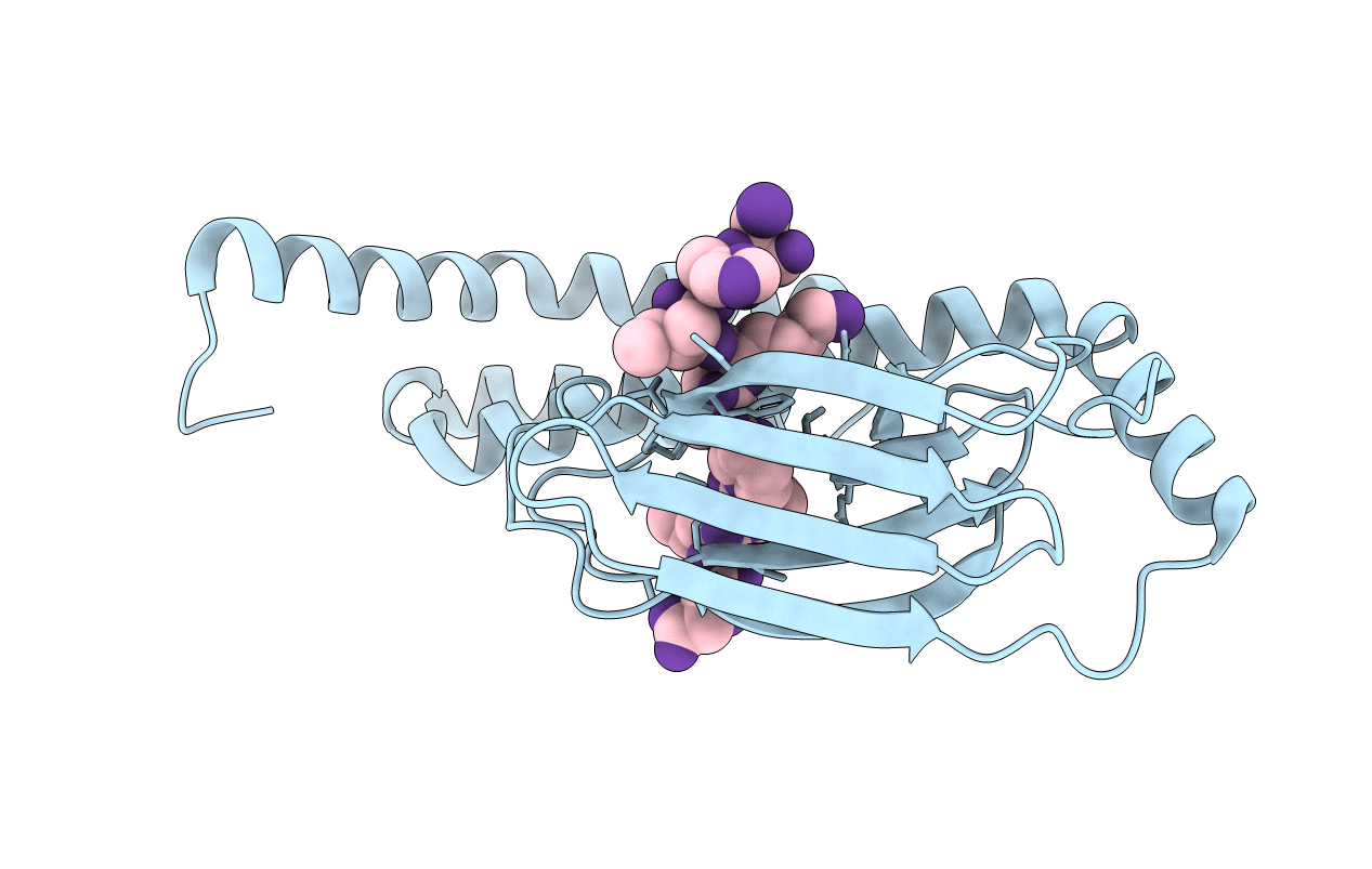
HscA substrate binding domain complexed with the IscU recognition peptide ELPPVKIHC
Biological Source:
Source Organism:
Escherichia coli (Taxon ID: 562)
Host Organism:
Method Details:
Experimental Method:
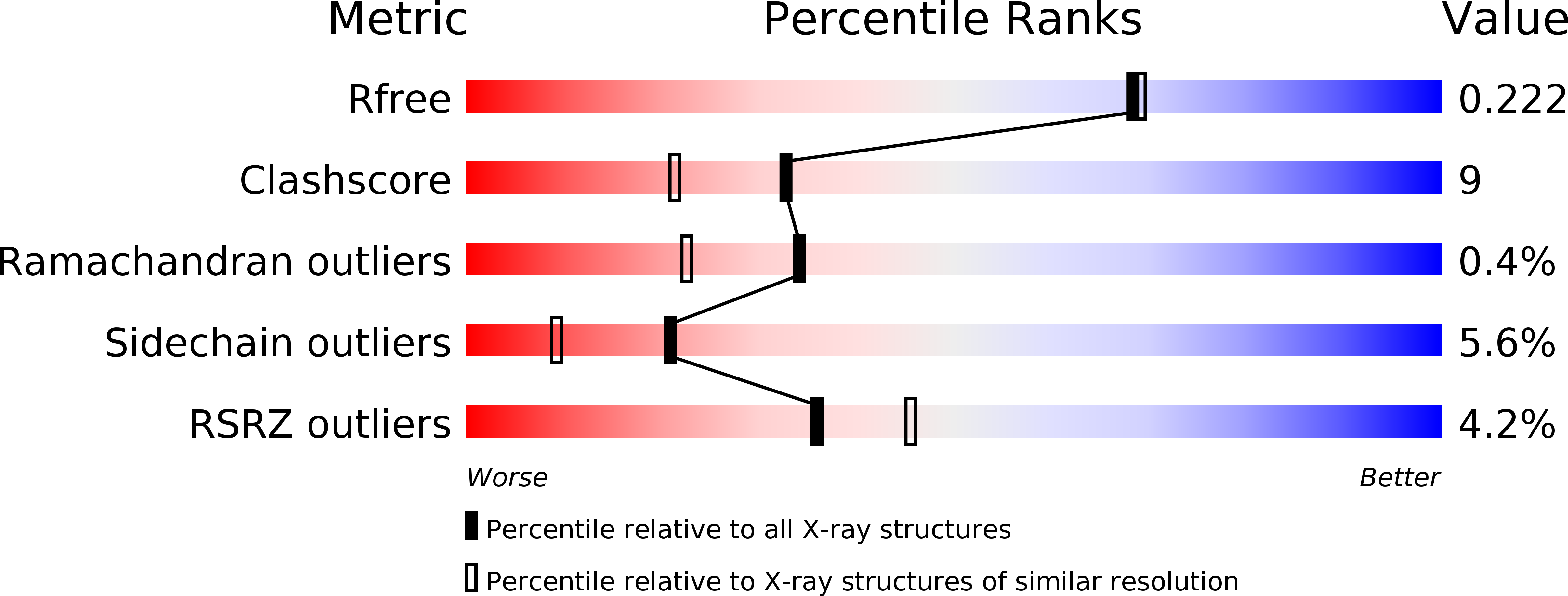
Resolution:
1.95 Å
R-Value Free:
0.21
R-Value Work:
0.17
R-Value Observed:
0.17
Space Group:
I 2 2 2
Macromolecular Entities
Structures with similar UniProt ID
Protein Blast
Polymer Type:polypeptide(L)
Molecule:Chaperone protein hscA
Gene (Uniprot):hscA
Chain IDs:A
Chain Length:227
Number of Molecules:1
Biological Source:Escherichia coli
Primary Citation
Crystal Structure of the Molecular Chaperone HscA Substrate Binding Domain Complexed with the IscU Recognition Peptide ELPPVKIHC.
J.Mol.Biol. 342 1265 1278 (2004)
PMID: 15351650 DOI: 10.1016/j.jmb.2004.07.025

Abstact

HscA, a specialized bacterial Hsp70-class molecular chaperone, interacts with the iron-sulfur cluster assembly protein IscU by recognizing a conserved LPPVK sequence motif. We report the crystal structure of the substrate-binding domain of HscA (SBD, residues 389-616) from Escherichia coli bound to an IscU-derived peptide, ELPPVKIHC. The crystals belong to the space group I222 and contain a single molecule in the asymmetric unit. Molecular replacement with the E.coli DnaK(SBD) model was used for phasing, and the HscA(SBD)-peptide model was refined to Rfactor=17.4% (Rfree=21.0%) at 1.95 A resolution. The overall structure of HscA(SBD) is similar to that of DnaK(SBD), although the alpha-helical subdomain (residues 506-613) is shifted up to 10 A relative to the beta-sandwich subdomain (residues 389-498) when compared to DnaK(SBD). The ELPPVKIHC peptide is bound in an extended conformation in a hydrophobic cleft in the beta-subdomain, which appears to be solvent-accessible via a narrow passageway between the alpha and beta-subdomains. The bound peptide is positioned in the reverse orientation of that observed in the DnaK(SBD)-NRLLLTG peptide complex placing the N and C termini of the peptide on opposite sides of the HscA(SBD) relative to the DnaK(SBD) complex. Modeling of the peptide in the DnaK-like forward orientation suggests that differences in hydrogen bonding interactions in the binding cleft and electrostatic interactions involving surface residues near the cleft contribute to the observed directional preference.

Legend

Protein

Chemical

Disease

Primary Citation of related structures
Feedback Form
Name
Email
Institute
Feedback