1TZI image
Deposition Date 2004-07-10
Release Date 2004-08-31
Last Version Date 2024-11-06
Entry Detail
PDB ID:
1TZI
Keywords:
Title:
Crystal Structure of the Fab YADS2 Complexed with h-VEGF
Biological Source:
Source Organism:
Mus musculus (Taxon ID: 10090)
Homo sapiens (Taxon ID: 9606)
Host Organism:
Method Details:
Experimental Method:
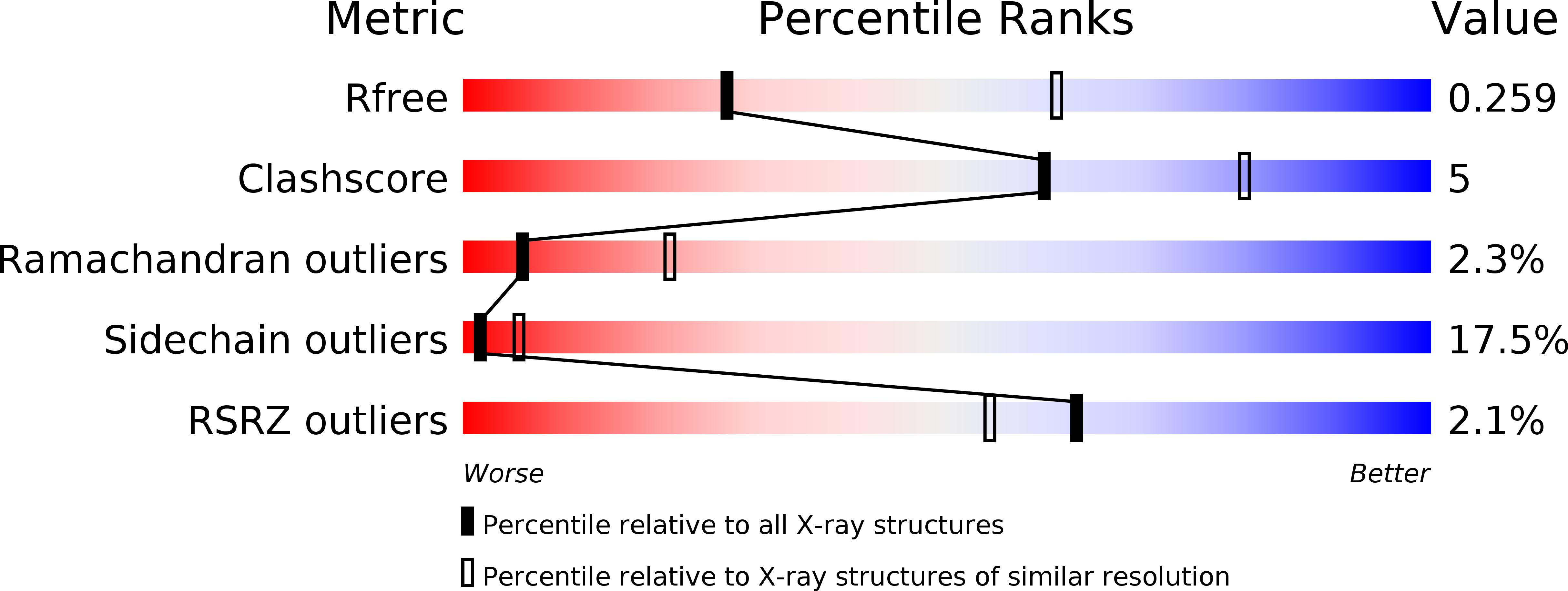
Resolution:
2.80 Å
R-Value Free:
0.25
R-Value Work:
0.21
R-Value Observed:
0.21
Space Group:
C 2 2 21
Macromolecular Entities
Polymer Type:polypeptide(L)
Molecule:Fab YADS2 Light Chain
Chain IDs:A
Chain Length:214
Number of Molecules:1
Biological Source:Mus musculus
Polymer Type:polypeptide(L)
Molecule:Fab YADS2 Heavy Chain
Chain IDs:B
Chain Length:222
Number of Molecules:1
Biological Source:Mus musculus
Structures with similar UniProt ID
Protein Blast
Polymer Type:polypeptide(L)
Molecule:Vascular endothelial growth factor A
Gene (Uniprot):VEGFA
Chain IDs:C (auth: V)
Chain Length:102
Number of Molecules:1
Biological Source:Homo sapiens
Ligand Molecules
Primary Citation
Synthetic antibodies from a four-amino-acid code: A dominant role for tyrosine in antigen recognition
Proc.Natl.Acad.Sci.USA 101 12467 12472 (2004)
PMID: 15306681 DOI: 10.1073/pnas.0401786101

Abstact

Antigen-binding fragments (Fabs) with synthetic antigen-binding sites were isolated from phage-displayed libraries with restricted complementarity-determining region (CDR) diversity. Libraries were constructed such that solvent-accessible CDR positions were randomized with a degenerate codon that encoded for only four amino acids (tyrosine, alanine, aspartate, and serine). Nonetheless, high-affinity Fabs (K(d) = 2-10 nM) were isolated against human vascular endothelial growth factor (hVEGF), and the crystal structures were determined for two distinct Fab-hVEGF complexes. The structures revealed that antigen recognition was mediated primarily by tyrosine side chains, which accounted for 71% of the Fab surface area that became buried upon binding to hVEGF. In contrast, aspartate residues within the CDRs were almost entirely excluded from the binding interface. Alanine and serine residues did not make many direct contacts with antigen, but they allowed for space and conformational flexibility and thus played an auxiliary role in facilitating productive contacts between tyrosine and antigen. Tyrosine side chains were capable of mediating most of the contacts necessary for high-affinity antigen recognition, and, thus, it seems likely that the overabundance of tyrosine in natural antigen-binding sites is a consequence of the side chain being particularly well suited for making productive contacts with antigen. The findings shed light on the basic principles governing the evolution of natural immune repertoires and should also aid the development of improved synthetic antibody libraries.

Legend

Protein

Chemical

Disease

Primary Citation of related structures