Deposition Date
2004-07-05
Release Date
2004-11-23
Last Version Date
2024-10-30
Entry Detail
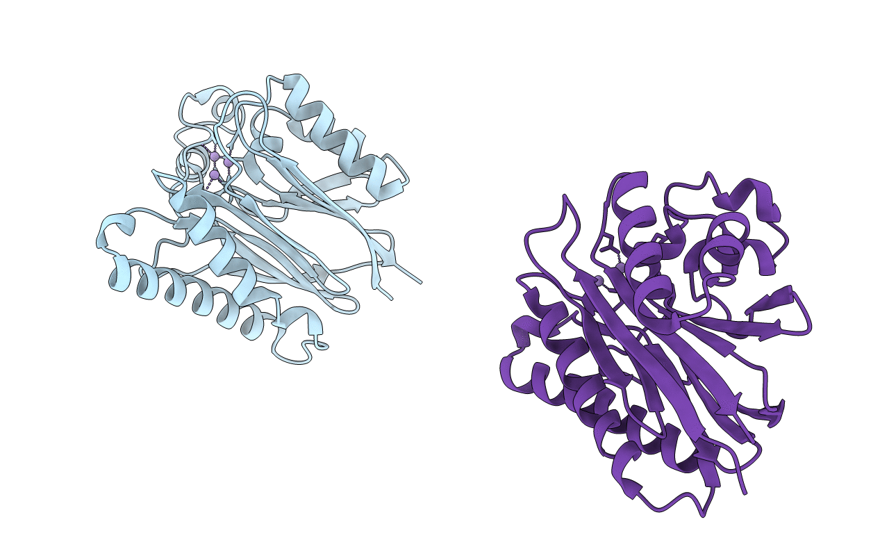
PDB ID:
1TXO
Keywords:
Title:
Crystal structure of the Mycobacterium tuberculosis serine/threonine phosphatase PstP/Ppp at 1.95 A.
Biological Source:
Source Organism(s):
Mycobacterium tuberculosis (Taxon ID: 1773)
Expression System(s):
Method Details:
Experimental Method:
Resolution:
1.95 Å
R-Value Free:
0.23
R-Value Work:
0.19
R-Value Observed:
0.20
Space Group:
P 1 21 1