1TW7 image
Deposition Date 2004-06-30
Release Date 2005-07-19
Last Version Date 2023-08-23
Entry Detail
PDB ID:
1TW7
Keywords:
Title:
Wide Open 1.3A Structure of a Multi-drug Resistant HIV-1 Protease Represents a Novel Drug Target
Biological Source:
Source Organism:
Method Details:
Experimental Method:
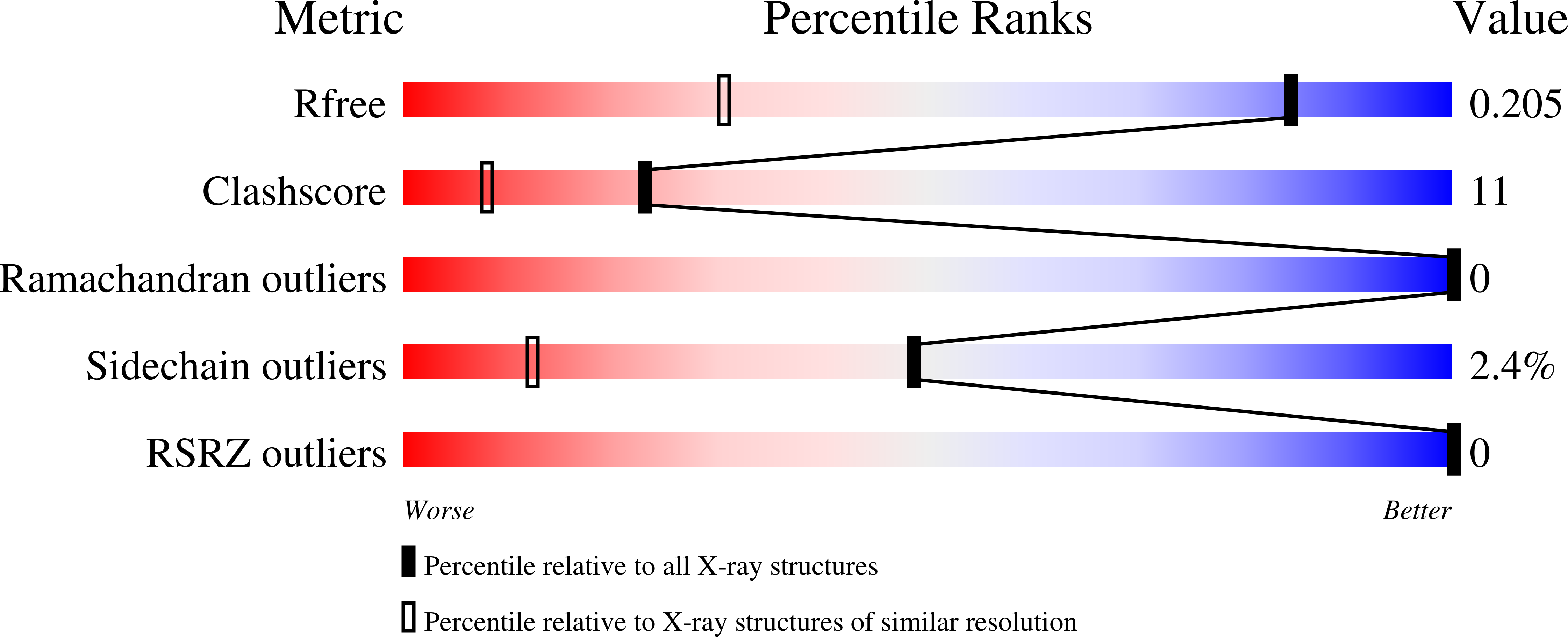
Resolution:
1.30 Å
R-Value Free:
0.21
R-Value Work:
0.14
R-Value Observed:
0.14
Space Group:
P 41
Macromolecular Entities
Polymer Type:polypeptide(L)
Molecule:protease
Mutations:L10I, D25N, M36V, M46L, I54V, I62V, L63P, A71V, V82A, I84V, L90M
Chain IDs:A, B
Chain Length:99
Number of Molecules:2
Biological Source:Human immunodeficiency virus 1
Ligand Molecules
Primary Citation

Abstact

This report examines structural changes in a highly mutated, clinical multidrug-resistant HIV-1 protease, and the crystal structure has been solved to 1.3 A resolution in the absence of any inhibitor. This protease variant contains codon mutations at positions 10, 36, 46, 54, 62, 63, 71, 82, 84, and 90 that confer resistance to protease inhibitors. Major differences between the wild-type and the variant include a structural change initiated by the M36V mutation and amplified by additional mutations in the flaps of the protease, resulting in a "wide-open" structure that represents an opening that is 8 A wider than the "open" structure of the wild-type protease. A second structural change is triggered by the L90M mutation that results in reshaping the 23-32 segment. A third key structural change of the protease is due to the mutations from longer to shorter amino acid side chains at positions 82 and 84.

Legend

Protein

Chemical

Disease

Primary Citation of related structures