Deposition Date
2004-06-30
Release Date
2005-05-17
Last Version Date
2023-10-25
Entry Detail
PDB ID:
1TVN
Keywords:
Title:
Cellulase cel5G from Pseudoalteromonas haloplanktis, A family GH 5-2 enzyme
Biological Source:
Source Organism(s):
Pseudoalteromonas haloplanktis (Taxon ID: 228)
Expression System(s):
Method Details:
Experimental Method:
Resolution:
1.41 Å
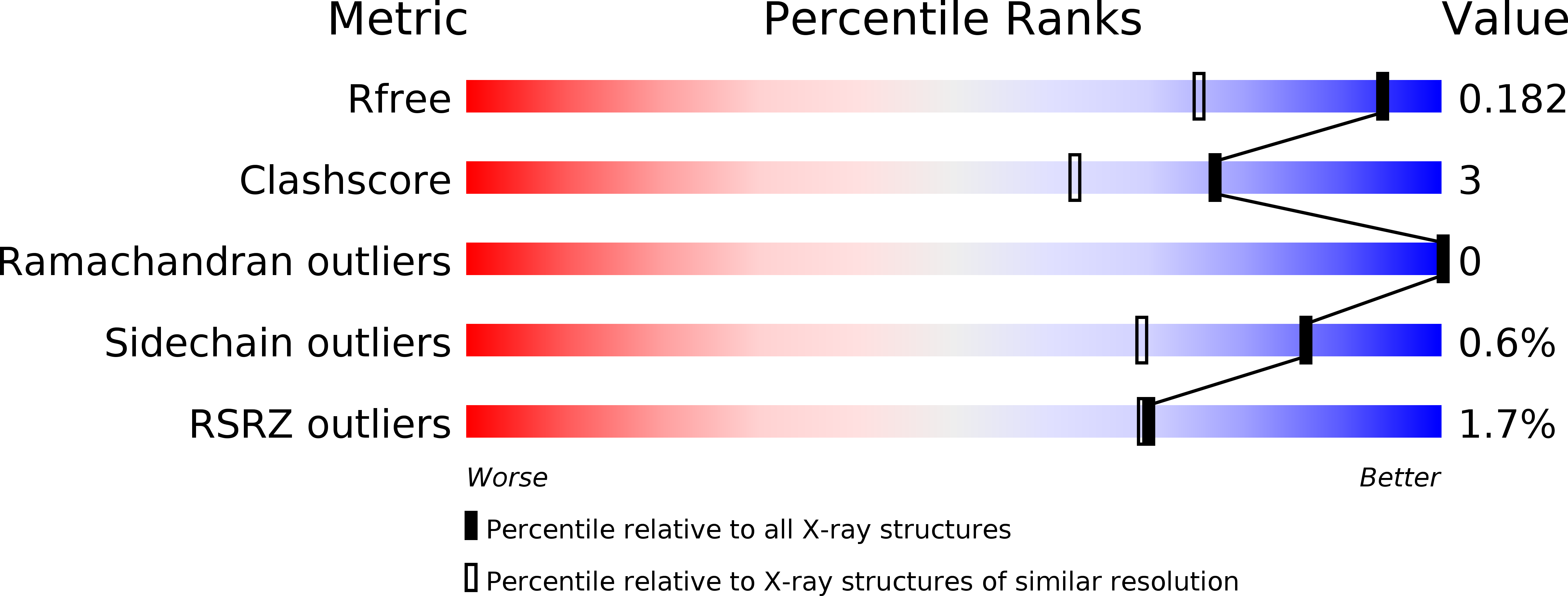
R-Value Free:
0.17
R-Value Work:
0.15
R-Value Observed:
0.15
Space Group:
P 21 21 21