1TV6 image
Entry Detail
PDB ID:
1TV6
Keywords:
Title:
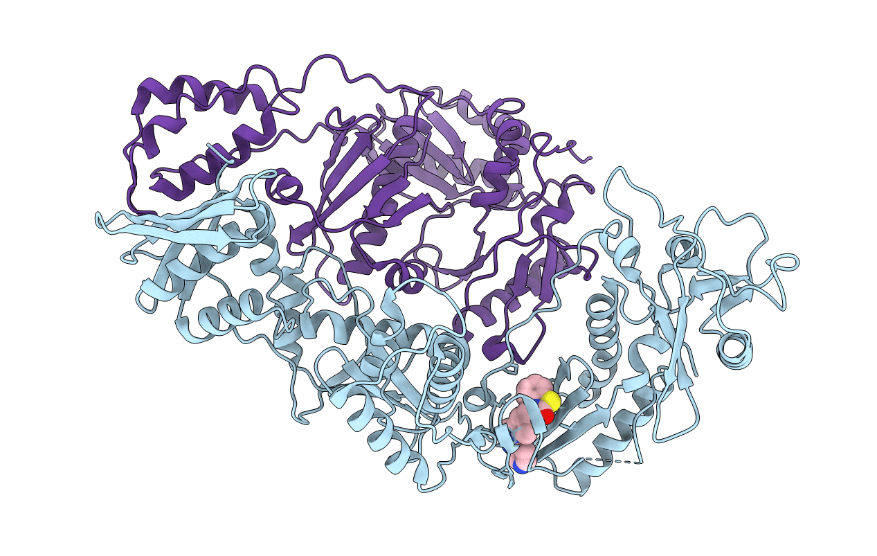
HIV-1 Reverse Transcriptase Complexed with CP-94,707
Biological Source:
PDB Version:
Deposition Date:
2004-06-28
Release Date:
2004-07-20
Method Details:
Experimental Method:
Resolution:
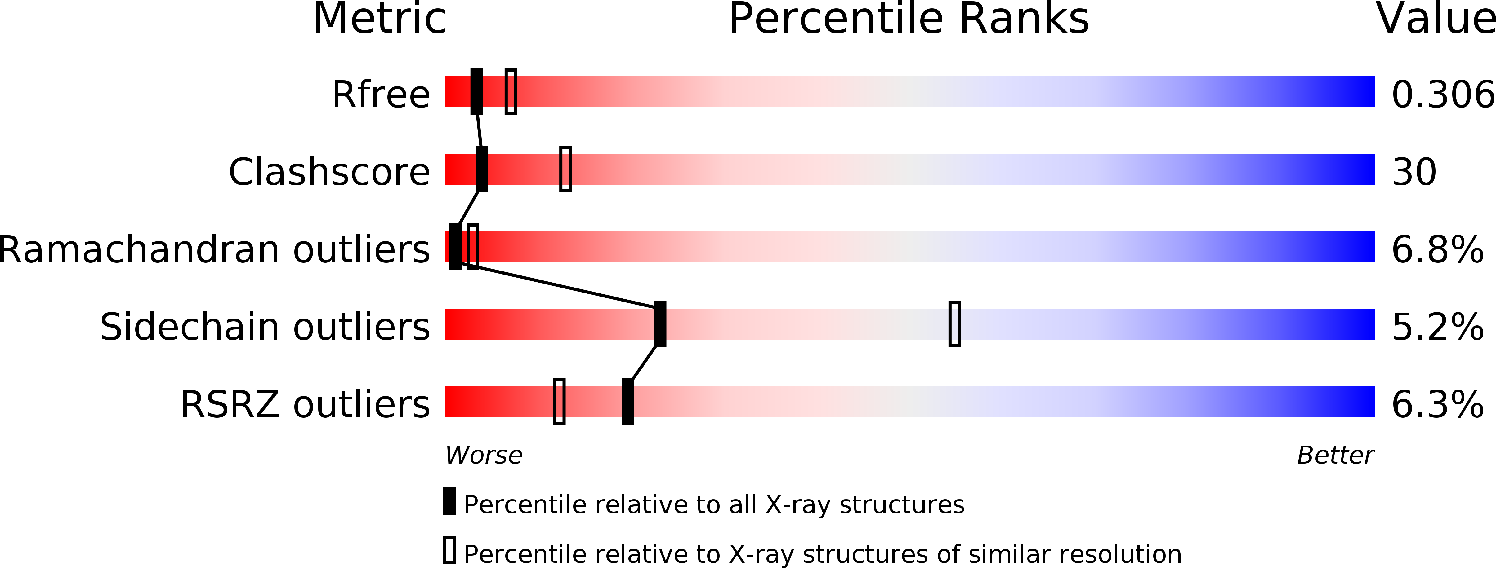
2.80 Å
R-Value Free:
0.31
R-Value Work:
0.26
R-Value Observed:
0.26
Space Group:
C 1 2 1
Macromolecular Entities
Polymer Type:polypeptide(L)
Description:reverse transcriptase p66 subunit
Chain IDs:A
Chain Length:560
Number of Molecules:1
Biological Source:Human immunodeficiency virus type 1 BH10
Polymer Type:polypeptide(L)
Description:reverse transcriptase p51 subunit
Chain IDs:B
Chain Length:440
Number of Molecules:1
Biological Source:Human immunodeficiency virus type 1 BH10
Ligand Molecules
Primary Citation
Structure of HIV-1 reverse transcriptase bound to an inhibitor active against mutant RTs resistant to other non-nucleoside inhibitors
Proc.Natl.Acad.Sci.USA 101 10548 10553 (2004)
PMID: 15249669 DOI: 10.1073/pnas.0404151101

Abstact

We have determined the crystal structure of the HIV type 1 reverse transcriptase complexed with CP-94,707, a new nonnucleoside reverse transcriptase inhibitor (NNRTI), to 2.8-A resolution. In addition to inhibiting the wild-type enzyme, this compound inhibits mutant enzymes that are resistant to inhibition by nevirapine, efavirenz, and delaviridine. In contrast to other NNRTI complexes where tyrosines 181 and 188 are pointing toward the enzyme active site, the binding pocket in this complex has the tyrosines pointing the opposite direction, as in the unliganded protein structure, to accommodate CP-94,707. This conformation of the pocket has not been observed previously in NNRTI complexes and substantially alters the shape and surface features that are available for interactions with the inhibitor. One ring of CP-94,707 makes extensive stacking interactions with tryptophan 229, one of the few residues in the NNRTI-binding pocket that cannot readily mutate to give rise to drug resistance. In this conformation of the pocket, mutations of tyrosines 181 and 188 are less likely to disrupt inhibitor binding. Modeling the asparagine mutation of lysine 103 shows that a hydrogen bond between it and tyrosine 188 could form as readily in the CP-94,707 complex as it does in the apoenzyme structure, providing an explanation for the activity of this inhibitor against this clinically important mutant.

Legend

Protein

Chemical

Disease

Primary Citation of related structures