1TUQ image
Deposition Date 2004-06-25
Release Date 2004-10-05
Last Version Date 2024-05-22
Entry Detail
PDB ID:
1TUQ
Keywords:
Title:
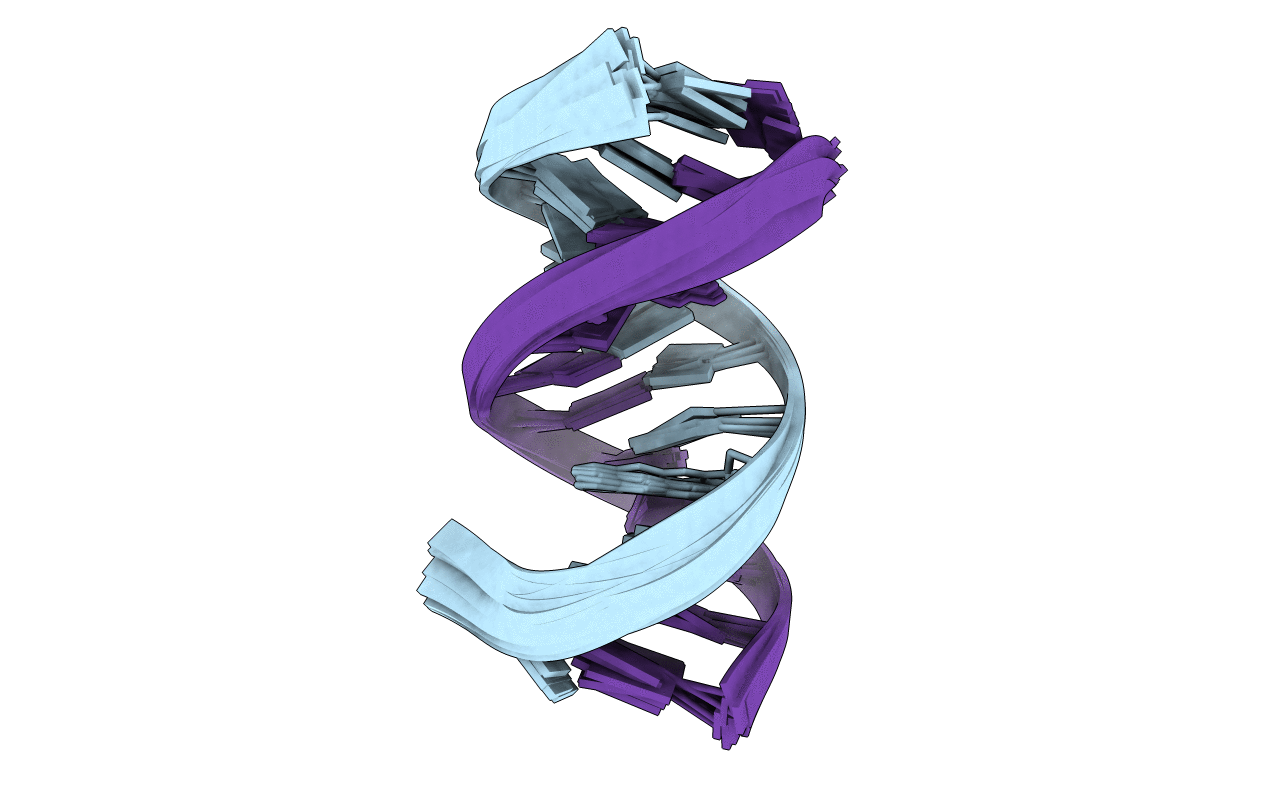
NMR Structure Analysis of the B-DNA Dodecamer CTCtCACGTGGAG with a tricyclic cytosin base analogue
Biological Source:
Source Organism:
(Taxon ID: ) (Taxon ID: )
Method Details:
Experimental Method:
Conformers Calculated:
100
Conformers Submitted:
10
Selection Criteria:
lowest CYANA target function
Macromolecular Entities
Polymer Type:polydeoxyribonucleotide
Molecule:5'-D(P*CP*TP*CP*(TC1)P*AP*CP*GP*TP*GP*GP*AP*G)-3'
Chain IDs:A, B
Chain Length:12
Number of Molecules:2
Biological Source:
Modified Residue
Compound ID Chain ID Parent Comp ID Details 2D Image
TC1 A DC ?
Ligand Molecules
Primary Citation
DNA adopts normal B-form upon incorporation of highly fluorescent DNA base analogue tC: NMR structure and UV-Vis spectroscopy characterization.
Nucleic Acids Res. 32 5087 5095 (2004)
PMID: 15452275 DOI: 10.1093/nar/gkh844

Abstact

The influence of the highly fluorescent tricyclic cytosine base analogue (tC) on duplex DNA conformation is investigated. The duplex properties are characterized by absorbance and circular dichroism (CD) for all combinations of neighbouring bases to tC, and an NMR structure is determined for one tC-containing sequence. For the oligonucleotides with one tC incorporated instead of cytosine, the melting temperature is increased on average by 2.7 degrees C above that for the unmodified ones. CD spectra are practically identical for modified and unmodified sequences, indicating an unperturbed B-DNA conformation. The NMR structure determination of the self-complementary sequence 5'-CTC(tC)ACGTGGAG shows a DNA conformation consistent with B-form for the whole duplex. The root-mean-square distance for the nucleotides of the eight central base pairs between the 10 structures with lowest CYANA target functions and a mean structure is 0.45 +/- 0.17 A. The NMR data confirm correct base pairing for tC by the observation of both intrastrand and interstrand imino proton NOEs. Altogether, this suggests that tC works well as a cytosine analogue, i.e. it is situated in the base stack, forming hydrogen bonds with G in the complementary strand, without distorting the DNA backbone conformation. This first example of an artificial, highly fluorescent DNA base that does not perturb the DNA conformation could have valuable applications for the study of the structure and dynamics of nucleic acid systems.

Legend

Protein

Chemical

Disease

Primary Citation of related structures