1TUG image
Deposition Date 2004-06-24
Release Date 2004-07-20
Last Version Date 2024-10-30
Entry Detail
PDB ID:
1TUG
Title:
Aspartate Transcarbamoylase Catalytic Chain Mutant E50A Complex with Phosphonoacetamide, Malonate, and Cytidine-5-Prime-Triphosphate (CTP)
Biological Source:
Source Organism:
Escherichia coli (Taxon ID: 562)
Host Organism:
Method Details:
Experimental Method:
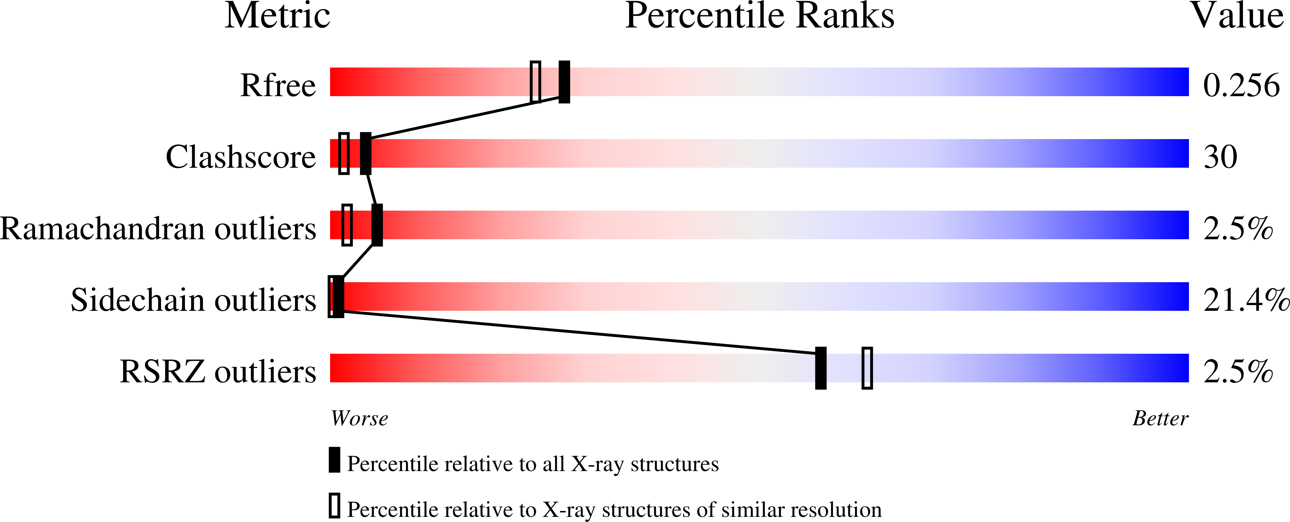
Resolution:
2.10 Å
R-Value Free:
0.27
R-Value Observed:
0.21
Space Group:
P 3 2 1
Macromolecular Entities
Polymer Type:polypeptide(L)
Molecule:Aspartate carbamoyltransferase catalytic chain
Gene (Uniprot):pyrB
Mutations:E50A
Chain IDs:A, C
Chain Length:310
Number of Molecules:2
Biological Source:Escherichia coli
Polymer Type:polypeptide(L)
Molecule:Aspartate carbamoyltransferase regulatory chain
Gene (Uniprot):pyrI
Chain IDs:B, D
Chain Length:153
Number of Molecules:2
Biological Source:Escherichia coli
Primary Citation
Monitoring the Transition from the T to the R State in E.coli Aspartate Transcarbamoylase by X-ray Crystallography: Crystal Structures of the E50A Mutant Enzyme in Four Distinct Allosteric States.
J.Mol.Biol. 341 853 868 (2004)
PMID: 15288791 DOI: 10.1016/j.jmb.2004.06.002

Abstact

A detailed description of the transition that allosteric enzymes undergo constitutes a major challenge in structural biology. We have succeeded in trapping four distinct allosteric states of a mutant enzyme of Escherichia coli aspartate transcarbomylase and determining their structures by X-ray crystallography. The mutant version of aspartate transcarbamoylase in which Glu50 in the catalytic chains was replaced by Ala destabilizes the native R state and shifts the equilibrium towards the T state. This behavior allowed the use of substrate analogs such as phosphonoacetamide and malonate to trap the enzyme in T-like and R-like structures that are distinct from the T-state structure of the wild-type enzyme (as represented by the structure of the enzyme with CTP bound and the R-state structure as represented by the structure with N-(phosphonacetyl)-L-aspartate bound). These structures shed light on the nature and the order of internal structural rearrangements during the transition from the T to the R state. They also suggest an explanation for diminished activity of the E50A enzyme and for the change in reaction mechanism from ordered to random for this mutant enzyme.

Legend

Protein

Chemical

Disease

Primary Citation of related structures